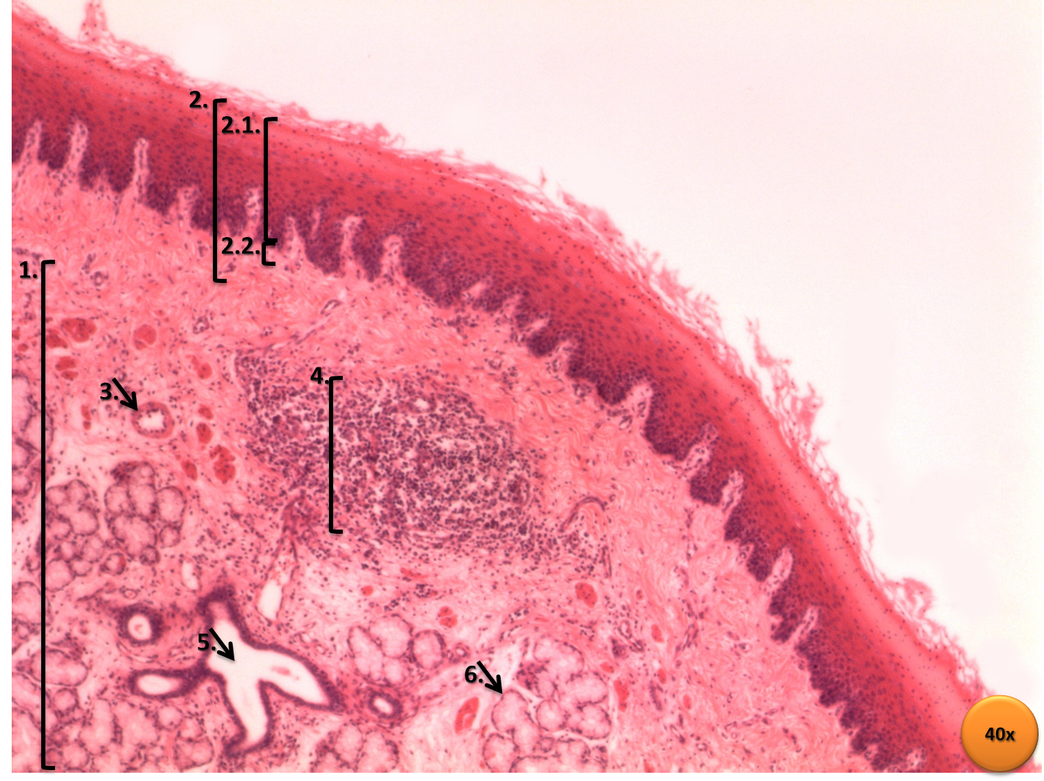
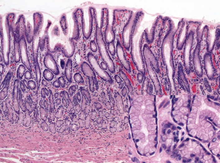
Normal·GI·Small intestine
Simple columnar epithelium — small intestine villus
Stain: H&E·Magnification: 40x·Tissue: Simple columnar with microvilli·4 labeled regions
Wikimedia Commons · File:Small intestine low mag.jpg · https://commons.wikimedia.org/wiki/File:Small_intestine_low_mag.jpg (CC-licensed)
Open the interactive viewer
Sign up free to pan/zoom up to 10×, tap each labeled marker for the structure's description and clinical context, and switch to quiz mode to test yourself on every region.
Create free accountDescription
Tall absorptive enterocytes line the villus surface. Apical brush border (microvilli) maximizes surface area; scattered goblet cells secrete mucin.
Labeled regions (4)
- 1Enterocyte (absorptive cell)
Tall columnar with eosinophilic apical border; expresses brush-border disaccharidases.
- 2Goblet cell
Mucin-filled apical cytoplasm; PAS-positive. Scattered among enterocytes.
- 3Brush border (microvilli)
Actin-based projections; site of disaccharidases (lactase first lost in injury).
- 4Lamina propria
Loose connective tissue core of villus; contains capillaries, lacteal, immune cells.