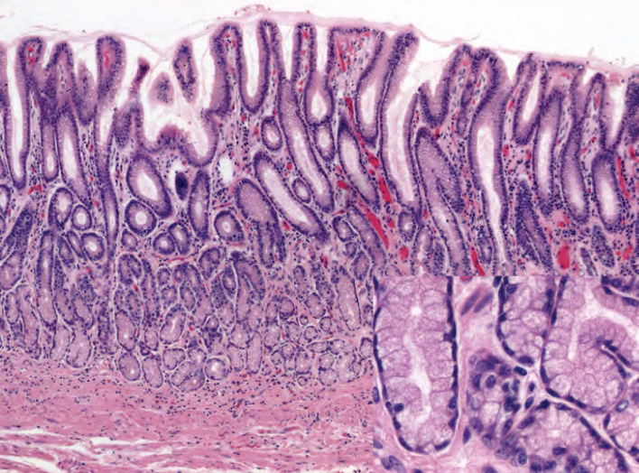
Normal·GI·Esophagus
Esophagus — non-keratinized stratified squamous epithelium
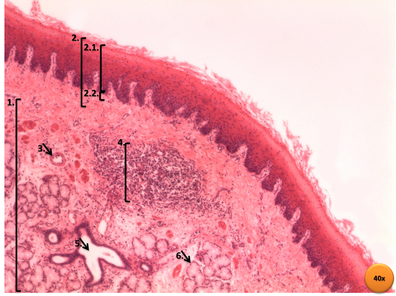
Stain: H&E·Magnification: 40x·Tissue: Stratified squamous epithelium, non-keratinized·3 labeled regions
Wikimedia Commons · File:Esôfago – HE – 40x.png · https://commons.wikimedia.org/wiki/File:Es%C3%B4fago_%E2%80%93_HE_%E2%80%93_40x.png (CC-licensed)
Open the interactive viewer
Sign up free to pan/zoom up to 10×, tap each labeled marker for the structure's description and clinical context, and switch to quiz mode to test yourself on every region.
Create free accountDescription
Tough, abrasion-resistant lining of the food conduit. Non-keratinized (unlike epidermis) — surface cells retain nuclei. Barrett esophagus = metaplasia to intestinal-type columnar epithelium from chronic acid reflux (↑ adenocarcinoma risk).
Labeled regions (3)
- 1Surface squames
Flat surface cells RETAIN nuclei (non-keratinized) — distinguishes from epidermis.
- 2Basal layer
Cuboidal mitotically active cells on basement membrane; replenish surface as cells slough.
- 3Lamina propria
Loose CT under epithelium with capillaries + immune cells. Site of esophageal varices in portal HTN.