Normal·GI·Salivary gland
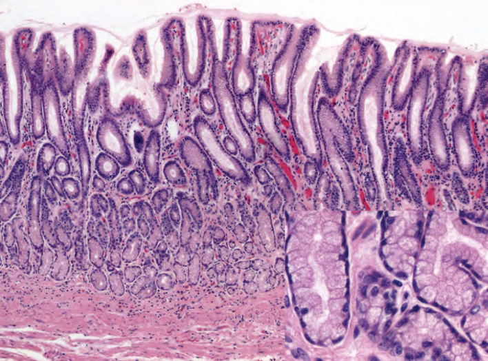
Submandibular salivary gland — mixed acini + serous demilunes
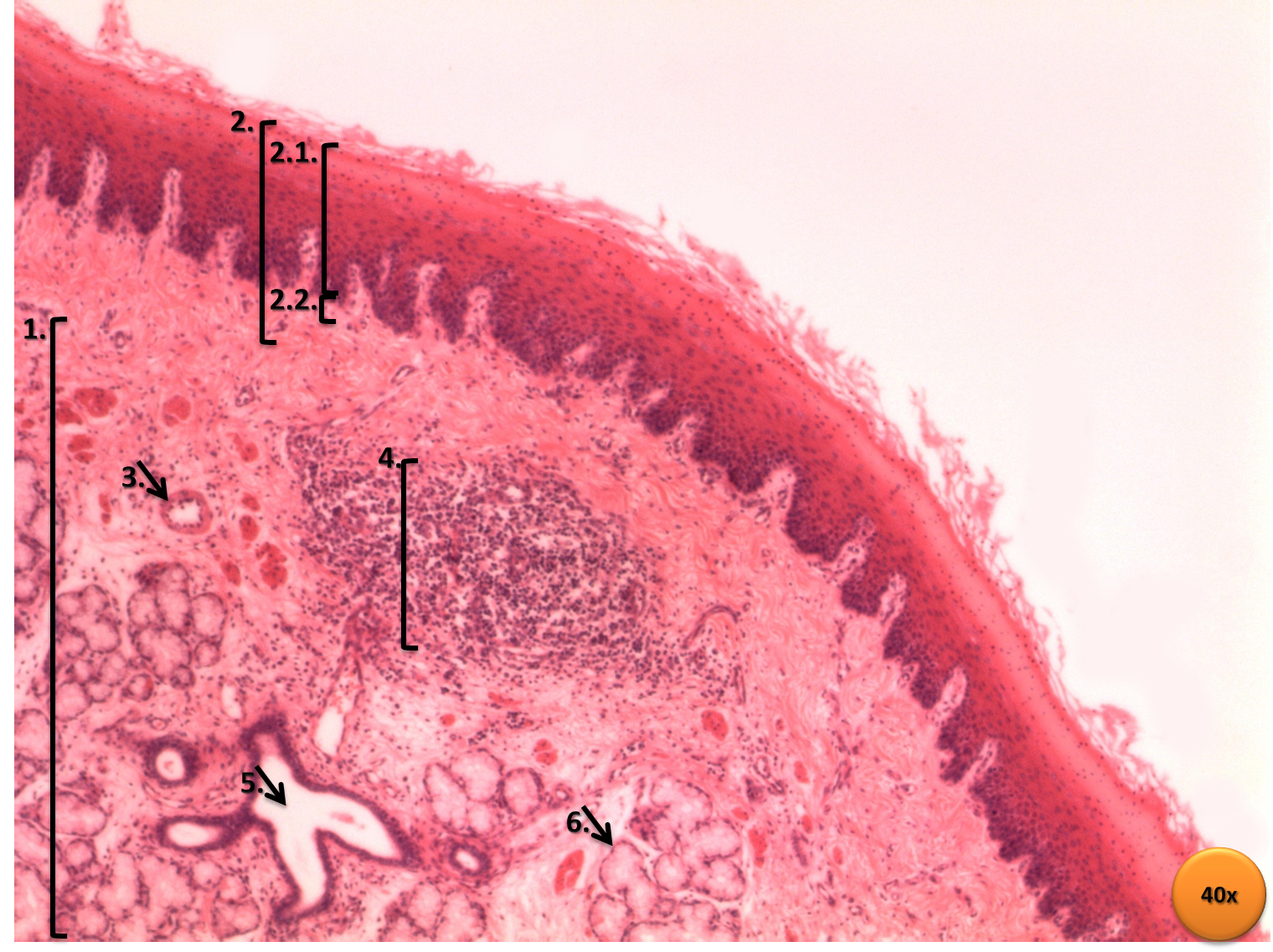
Stain: H&E·Magnification: 40x·Tissue: Mixed exocrine acini·3 labeled regions
Wikimedia Commons · File:Serous_demilunes_of_submandibular_gland_section.png · https://commons.wikimedia.org/wiki/File:Serous_demilunes_of_submandibular_gland_section.png (CC-licensed)
Open the interactive viewer
Sign up free to pan/zoom up to 10×, tap each labeled marker for the structure's description and clinical context, and switch to quiz mode to test yourself on every region.
Create free accountDescription
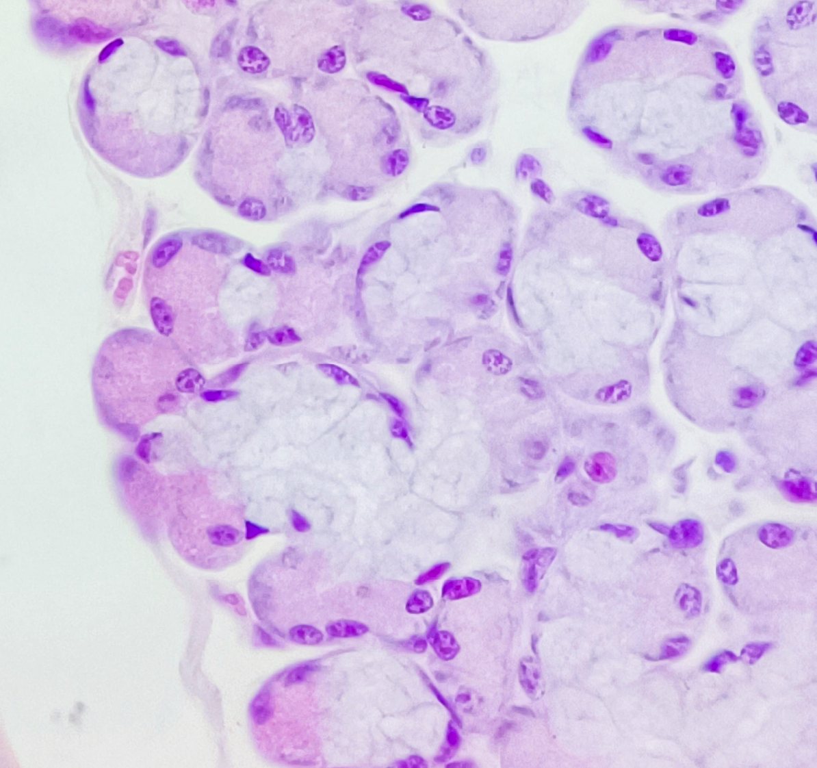
Mixed gland: pale mucous acini (mucin-filled cytoplasm) capped by darker serous demilunes (enzymatic secretions). Parotid = mostly serous; sublingual = mostly mucous; submandibular = mixed. Pleomorphic adenoma is most common salivary tumor.
Labeled regions (3)
- 1Mucous acinus
Pale, foamy cytoplasm with basally displaced flattened nuclei. Secretes mucin (lubrication).
- 2Serous demilune
Dark crescent of serous cells "capping" mucous acini. Secretes lysozyme + α-amylase + IgA.
- 3Striated duct
Cuboidal/columnar epithelium with basal striations (mitochondria-rich); modifies saliva by ion exchange.