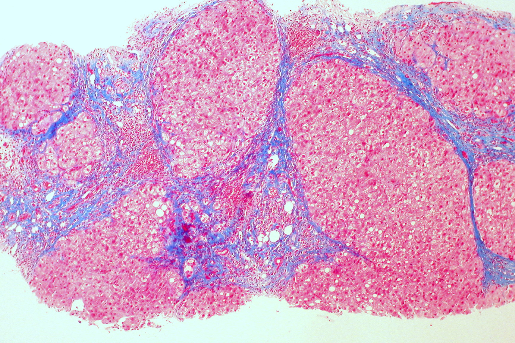
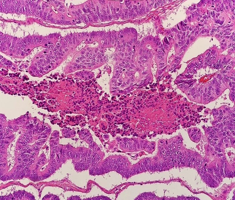
Pathologic·Pathology·Liver
Liver cirrhosis — regenerative nodules + bridging fibrosis (Trichrome)
Stain: Masson trichrome·Magnification: 4x·Tissue: Hepatic parenchyma with fibrosis·3 labeled regions
Wikimedia Commons · File:Cirrhosis_of_the_liver_(trichrome_stain)_(5690946257).jpg · https://commons.wikimedia.org/wiki/File:Cirrhosis_of_the_liver_(trichrome_stain)_(5690946257).jpg (CC-licensed)
Open the interactive viewer
Sign up free to pan/zoom up to 10×, tap each labeled marker for the structure's description and clinical context, and switch to quiz mode to test yourself on every region.
Create free accountDescription
End-stage chronic liver injury: parenchyma reorganized into regenerative nodules walled off by blue-staining (collagen, on Trichrome) bridging fibrous septa. Loss of normal lobular architecture → portal hypertension + impaired function.
Labeled regions (3)
- 1Regenerative nodule
Round island of hepatocytes that has regrown without normal lobular orientation. Lacks central vein.
- 2Bridging fibrosis (blue)
Bands of collagen (stellate cell-derived) connecting portal-portal or portal-central. Trichrome stains collagen blue.
- 3Distorted vascular architecture
Intrahepatic shunts across fibrous septa raise portal pressure → varices, ascites, splenomegaly.
More pathology histology
Comedo DCIS — high-grade ductal carcinoma in situ
Breast
Cervical intraepithelial neoplasia (CIN 2 / HSIL)
Cervix
Acute MI — coagulative necrosis of cardiomyocytes
Heart (myocardium)
Adenocarcinoma — colon
Colon
Granulomatous inflammation — non-caseating (sarcoid pattern)

Squamous cell carcinoma with keratin pearls