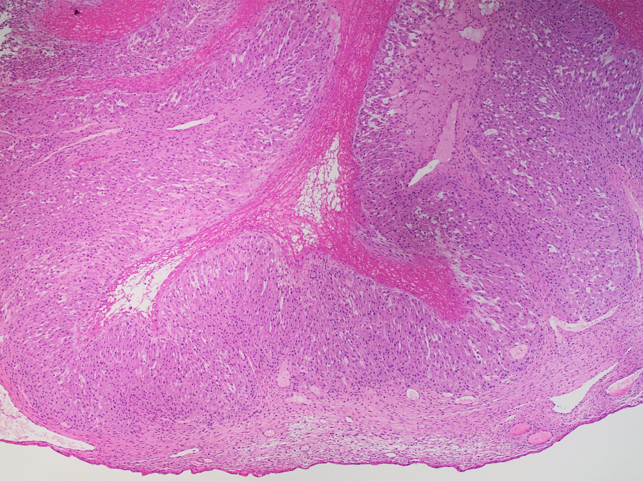
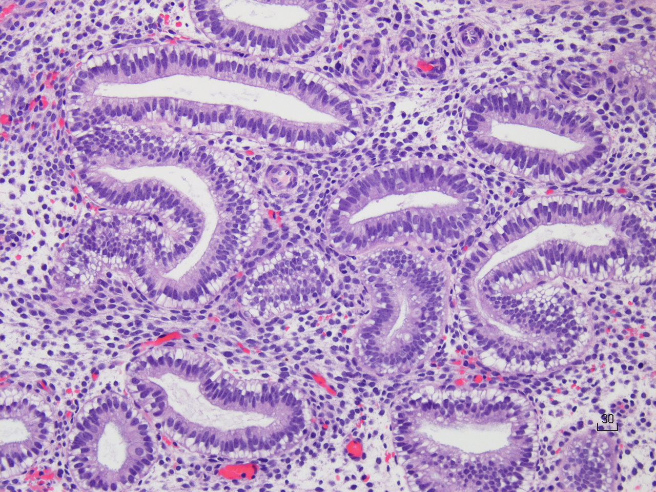
Normal·Reproductive·Testis
Testis — seminiferous tubules with spermatogenesis
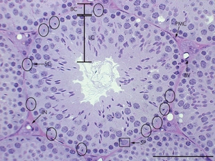
Stain: H&E·Magnification: 40x·Tissue: Seminiferous epithelium·5 labeled regions
Wikimedia Commons · File:Histological_structure_of_seminiferous_tubules_in_the_adult_mouse_testes..jpg · https://commons.wikimedia.org/wiki/File:Histological_structure_of_seminiferous_tubules_in_the_adult_mouse_testes..jpg (CC-licensed)
Open the interactive viewer
Sign up free to pan/zoom up to 10×, tap each labeled marker for the structure's description and clinical context, and switch to quiz mode to test yourself on every region.
Create free accountDescription
Coiled tubules lined by stratified seminiferous epithelium. Most basal cells = spermatogonia; progressively toward the lumen = primary spermatocytes (largest, leptotene/pachytene chromatin) → spermatids → spermatozoa. Interstitium contains Leydig cells.
Labeled regions (5)
- 1Spermatogonia
Basal layer stem cells (type A + B). Resting on basement membrane; replenish the germ line.
- 2Primary spermatocyte
Largest germ cell; characteristic clumped pachytene chromatin (looks "stringy"). Undergoing meiosis I.
- 3Spermatids / spermatozoa
Luminal-side; condensed dark heads + visible tails as they mature. Released into the lumen.
- 4Sertoli cell
Tall pale columnar cells spanning epithelium; form blood-testis barrier, secrete inhibin + androgen-binding protein. Tumor = Sertoli cell tumor.
- 5Leydig cell (interstitial)
Polygonal eosinophilic cells between tubules; secrete testosterone in response to LH. Contain lipid droplets + Reinke crystals.