Normal·Reproductive·Ovary
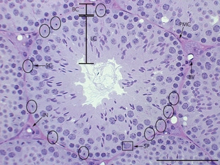
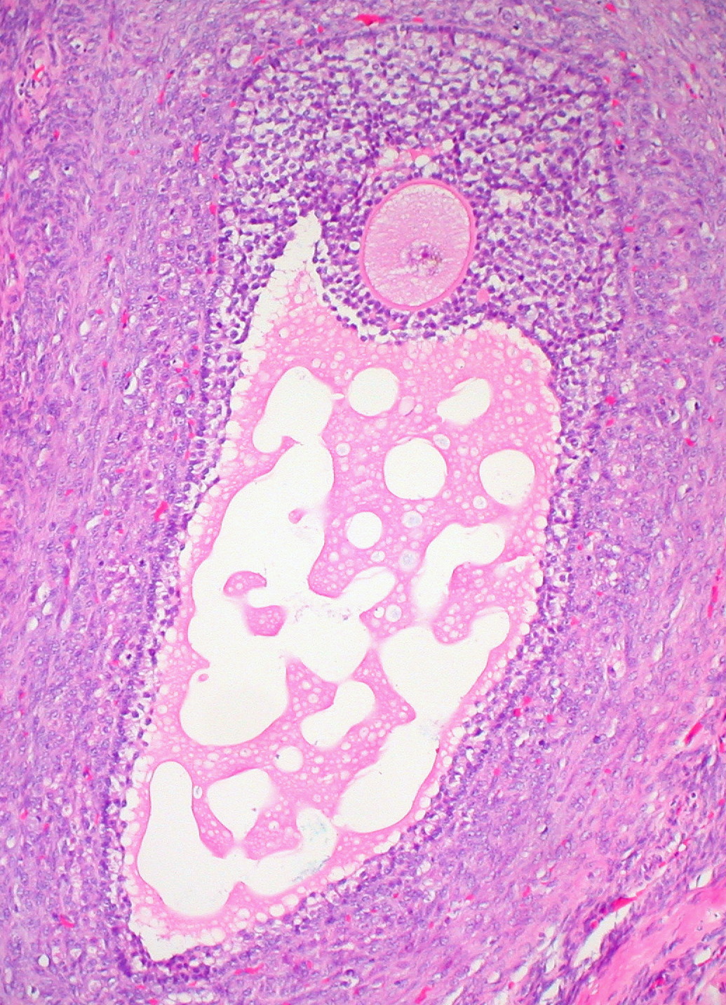
Ovary — Graafian (mature) follicle
Stain: H&E·Magnification: 10x·Tissue: Ovarian cortex with mature follicle·4 labeled regions
Wikimedia Commons · File:Graafian_Follicle,_Human_Ovary_(3595817584).jpg · https://commons.wikimedia.org/wiki/File:Graafian_Follicle,_Human_Ovary_(3595817584).jpg (CC-licensed)
Open the interactive viewer
Sign up free to pan/zoom up to 10×, tap each labeled marker for the structure's description and clinical context, and switch to quiz mode to test yourself on every region.
Create free accountDescription
Mature (Graafian) follicle just before ovulation: oocyte suspended in a large antrum, surrounded by cumulus oophorus + corona radiata, walled by granulosa + theca layers.
Labeled regions (4)
- 1Oocyte
Arrested in meiosis II until fertilization. Large eosinophilic cytoplasm with prominent nucleolus (germinal vesicle).
- 2Antrum
Fluid-filled cavity (follicular fluid). Defines the antral / Graafian stages.
- 3Granulosa layer
Stratified epithelial cells producing estrogen (aromatase, FSH-driven). Convert to corpus luteum after ovulation.
- 4Theca interna
Outer wall, vascularized stromal cells; LH-driven androgen production (substrate for granulosa aromatase).