Normal·Reproductive·Placenta
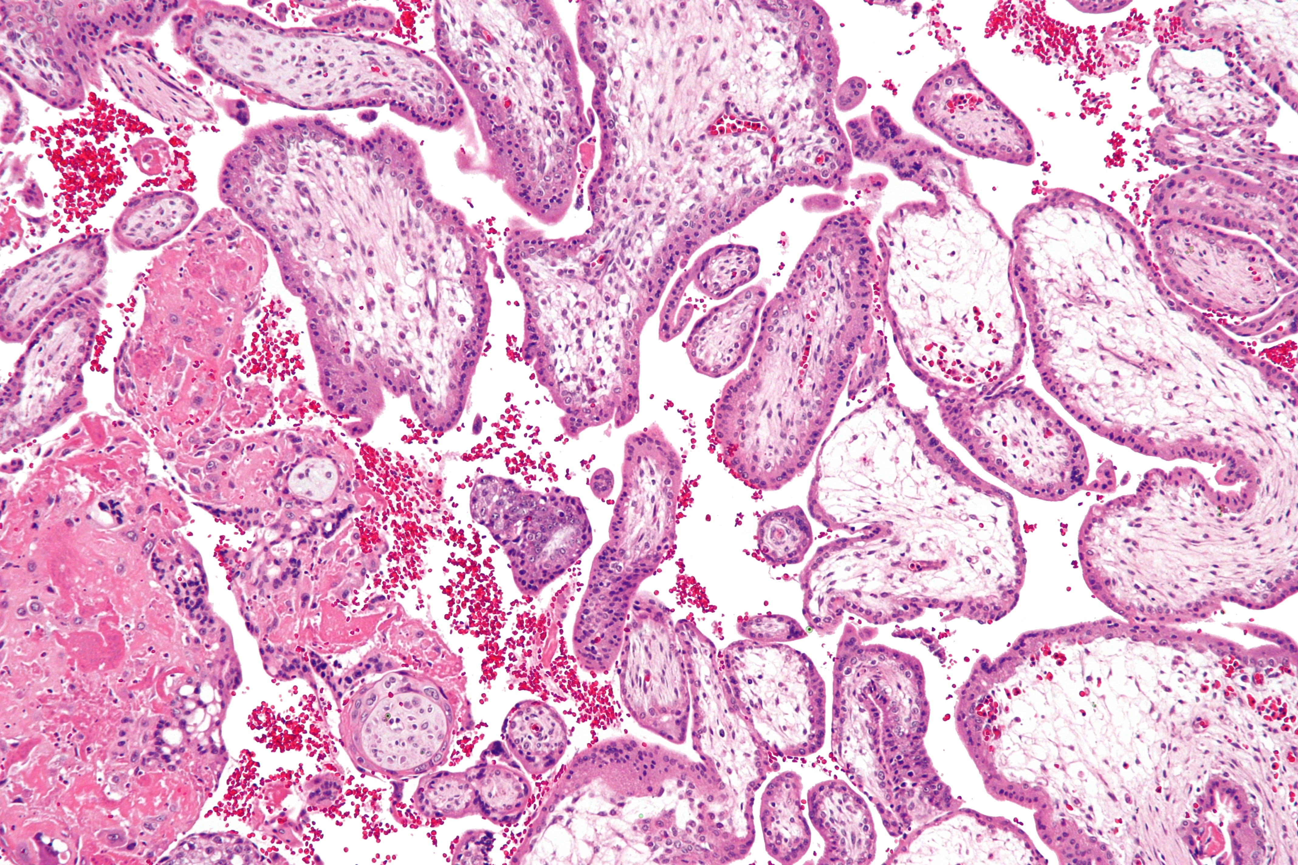
Placenta — chorionic villi with syncytiotrophoblast
Stain: H&E·Magnification: 20x·Tissue: Chorionic villus·4 labeled regions
Wikimedia Commons · File:Chorionic_villi_-_intermed_mag.jpg · https://commons.wikimedia.org/wiki/File:Chorionic_villi_-_intermed_mag.jpg (CC-licensed)
Open the interactive viewer
Sign up free to pan/zoom up to 10×, tap each labeled marker for the structure's description and clinical context, and switch to quiz mode to test yourself on every region.
Create free accountDescription
Fetal villi bathed in maternal blood within intervillous space. Two trophoblast layers (cytotrophoblast inner, syncytiotrophoblast outer) thin as pregnancy advances to facilitate gas/nutrient exchange. Syncytiotrophoblast secretes hCG, hPL, estrogen, progesterone.
Labeled regions (4)
- 1Syncytiotrophoblast
Outer multinucleated layer covering villus surface. Secretes β-hCG (basis of pregnancy test) + hPL + steroids.
- 2Cytotrophoblast (Langhans)
Inner mononuclear stem-cell layer beneath syncytium. Thins / fragments by third trimester. Source of choriocarcinoma.
- 3Fetal capillary
Within villus core — carries fetal blood (Hb F). Maternal-fetal exchange occurs across the trophoblast layers.
- 4Intervillous space
Bathed in maternal blood from spiral arteries. Drug + IgG transfer + gas/nutrient exchange occurs here.