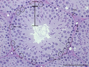
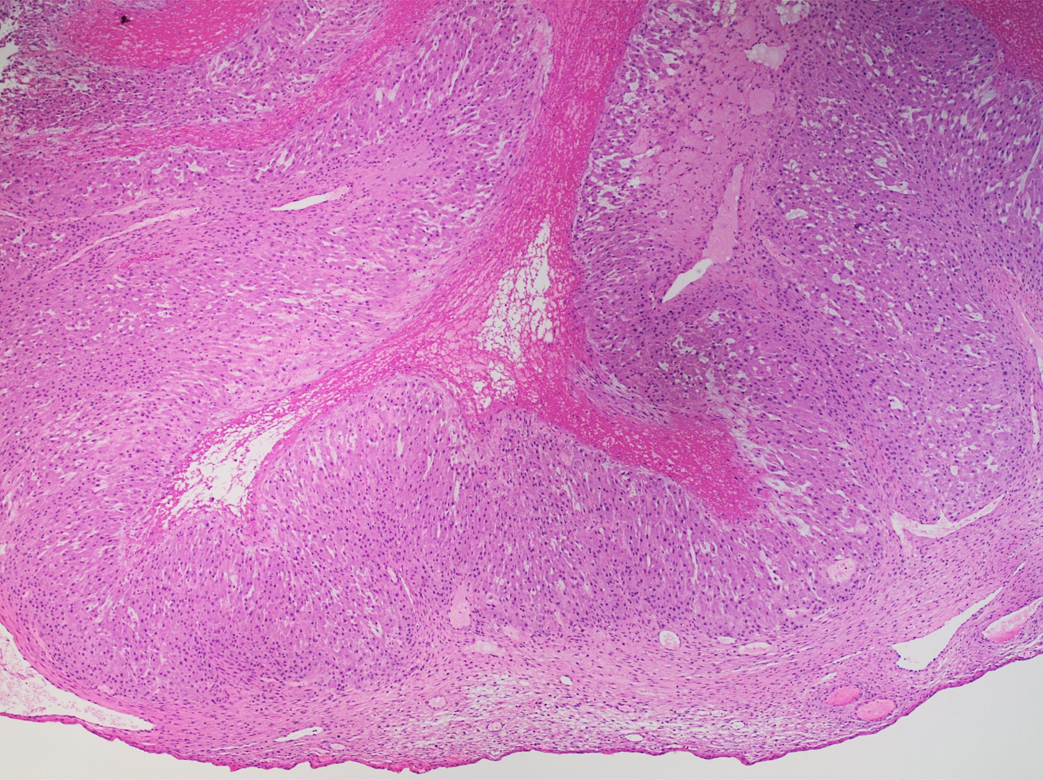
Normal·Reproductive·Ovary
Corpus luteum — luteinized granulosa + theca cells
Stain: H&E·Magnification: 10x·Tissue: Post-ovulatory follicle·3 labeled regions
Wikimedia Commons · File:Histopathology_of_a_corpus_luteum_cyst,_low_magnification.jpg · https://commons.wikimedia.org/wiki/File:Histopathology_of_a_corpus_luteum_cyst,_low_magnification.jpg (CC-licensed)
Open the interactive viewer
Sign up free to pan/zoom up to 10×, tap each labeled marker for the structure's description and clinical context, and switch to quiz mode to test yourself on every region.
Create free accountDescription
After ovulation, granulosa + theca cells "luteinize" — enlarge, fill with lipid + lutein pigment, and secrete progesterone (sustains endometrium). Regresses to corpus albicans without pregnancy; persists as corpus luteum of pregnancy under hCG.
Labeled regions (3)
- 1Luteinized granulosa cells
Large polygonal cells with abundant pale eosinophilic cytoplasm — secrete progesterone + some estrogen.
- 2Luteinized theca cells
Smaller, more deeply staining cells at the periphery; produce androgens converted to estrogen by granulosa aromatase.
- 3Central cavity
Site of prior ruptured follicle / hemorrhage. Becomes fibrotic corpus albicans without pregnancy.