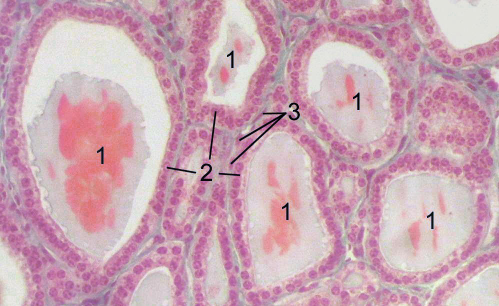
Normal·Endocrine·Thyroid
Thyroid — follicles + colloid
Stain: H&E·Magnification: 20x·Tissue: Follicular epithelium·3 labeled regions
Wikimedia Commons · File:Thyroid-histology.jpg · https://commons.wikimedia.org/wiki/File:Thyroid-histology.jpg (CC-licensed)
Open the interactive viewer
Sign up free to pan/zoom up to 10×, tap each labeled marker for the structure's description and clinical context, and switch to quiz mode to test yourself on every region.
Create free accountDescription
Single-layered cuboidal follicular cells surround a colloid-filled lumen storing thyroglobulin. C-cells (parafollicular) lie between follicles, producing calcitonin.
Labeled regions (3)
- 1Follicular cell
Cuboidal in normal state; becomes columnar when stimulated.
- 2Colloid
Eosinophilic thyroglobulin store.
- 3Parafollicular (C) cell
Larger, pale cytoplasm; produces calcitonin; origin of medullary thyroid carcinoma.