Normal·Endocrine·Parathyroid gland
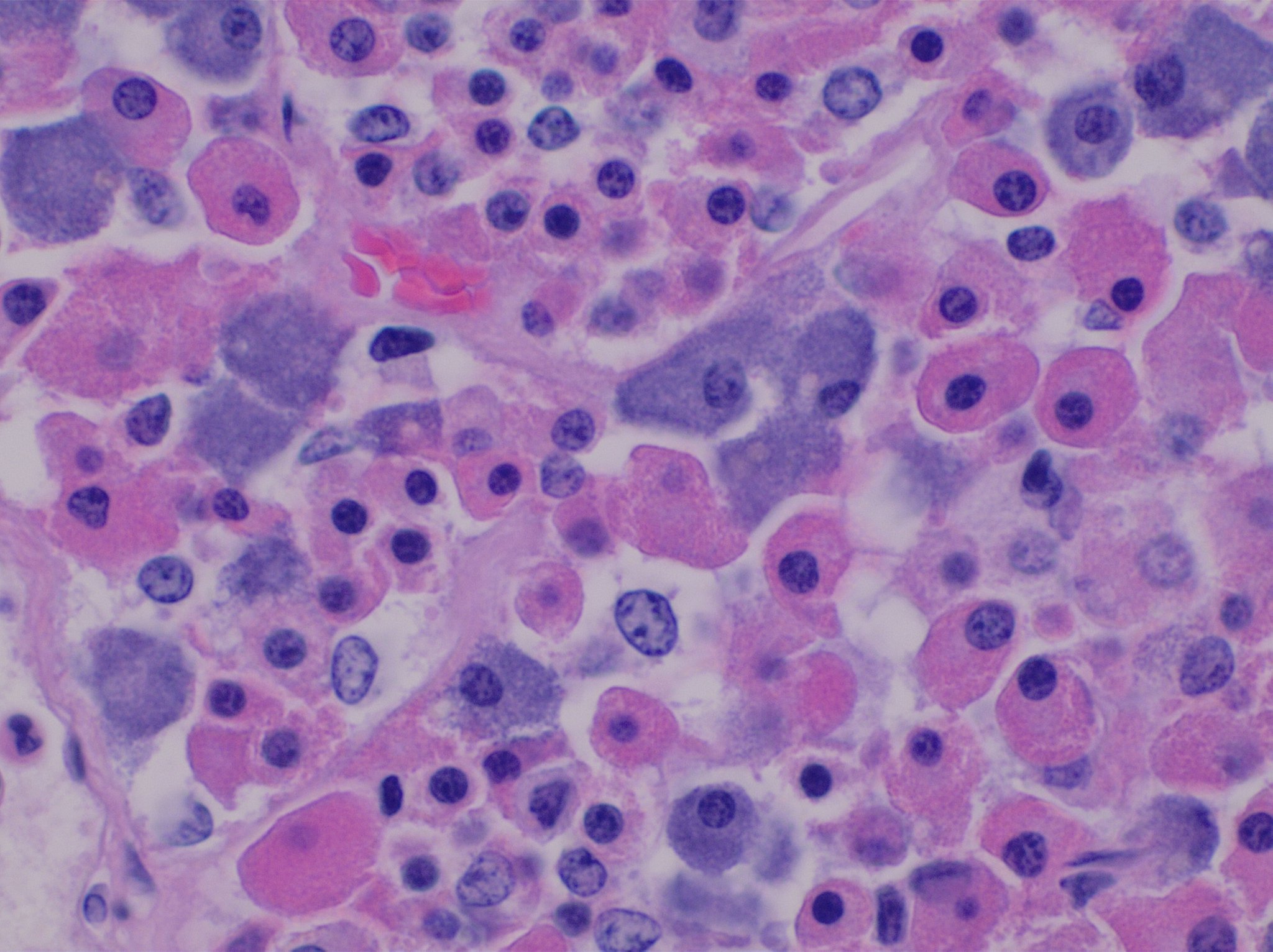
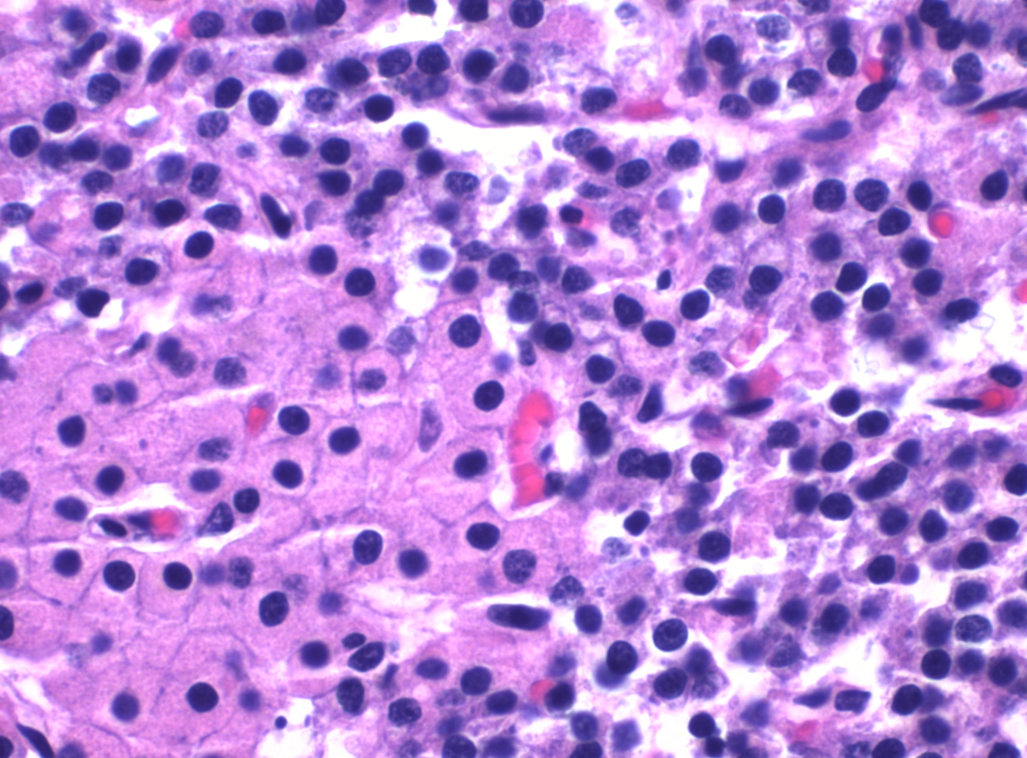
Parathyroid — chief + oxyphil cells
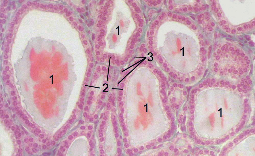
Stain: H&E·Magnification: 40x·Tissue: Endocrine parenchyma·3 labeled regions
Wikimedia Commons · File:Parathyroid_oxyphil_and_chief_cells.jpg · https://commons.wikimedia.org/wiki/File:Parathyroid_oxyphil_and_chief_cells.jpg (CC-licensed)
Open the interactive viewer
Sign up free to pan/zoom up to 10×, tap each labeled marker for the structure's description and clinical context, and switch to quiz mode to test yourself on every region.
Create free accountDescription
Two endocrine cell populations: small basophilic chief cells (secrete PTH; respond to ionized Ca²⁺ via CaSR) and larger eosinophilic oxyphil cells (mitochondria-rich, function unclear). PTH ↑ bone resorption + ↑ renal Ca reabsorption + ↑ calcitriol production.
Labeled regions (3)
- 1Chief cell
Small, round, basophilic; secretes parathyroid hormone (PTH). Hyperplastic in primary hyperparathyroidism + secondary HPT (CKD).
- 2Oxyphil cell
Larger, eosinophilic, mitochondria-packed. Increase with age; function unclear. Source of some parathyroid adenomas.
- 3Adipose tissue
Increases with age — by adulthood ~50% of gland is fat. Adenomas displace fat → uniform cellular field.