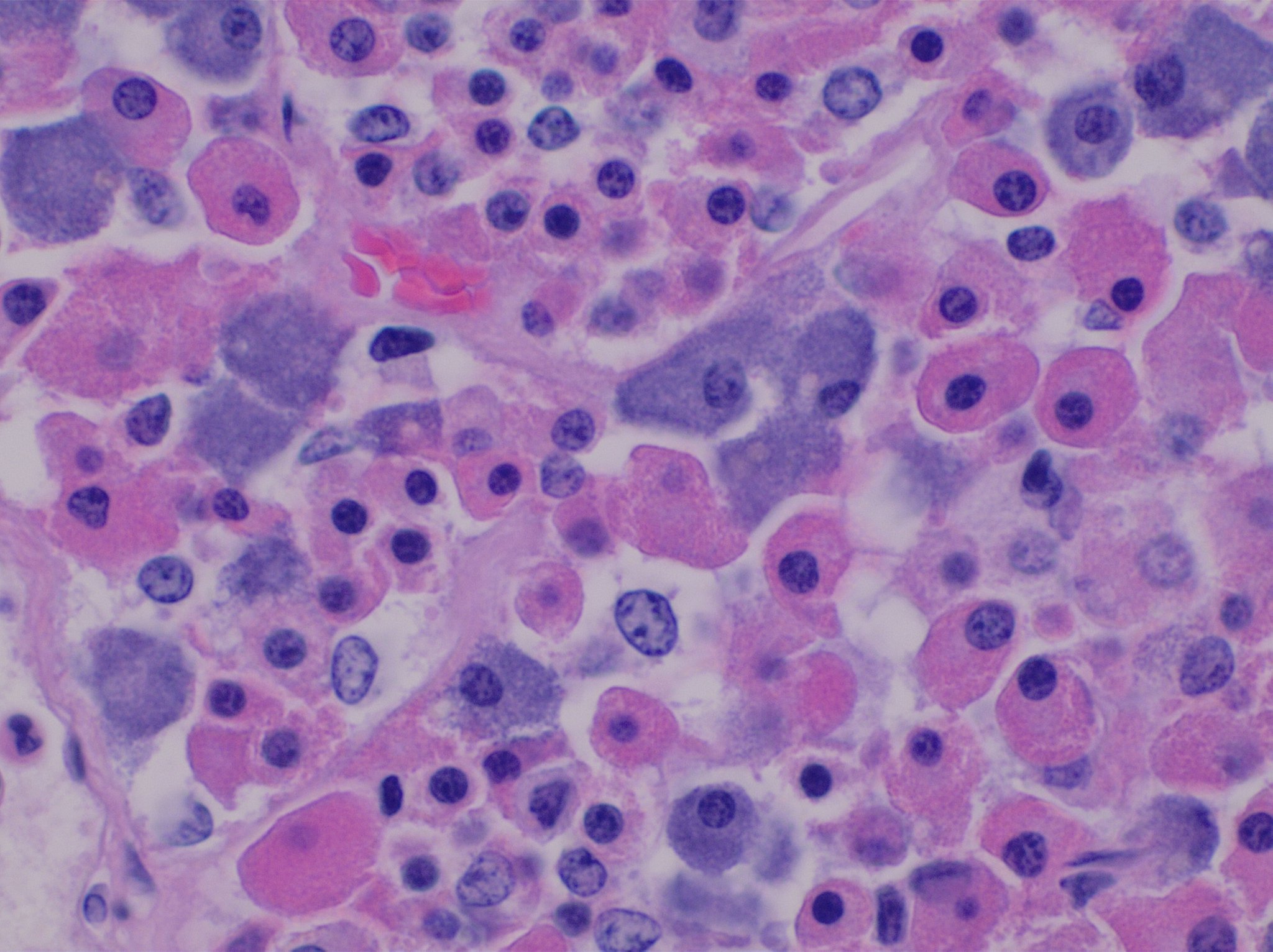
Normal·Endocrine·Pituitary gland
Anterior pituitary — acidophils, basophils, chromophobes
Stain: H&E·Magnification: 40x·Tissue: Pars distalis·3 labeled regions
Wikimedia Commons · File:Histology_of_pars_distalis_of_the_anterior_pituitary_with_chromophobes,_basophils,_and_acidophils.jpg · https://commons.wikimedia.org/wiki/File:Histology_of_pars_distalis_of_the_anterior_pituitary_with_chromophobes,_basophils,_and_acidophils.jpg (CC-licensed)
Open the interactive viewer
Sign up free to pan/zoom up to 10×, tap each labeled marker for the structure's description and clinical context, and switch to quiz mode to test yourself on every region.
Create free accountDescription
Three classic Romanowsky-staining classes: acidophils (pink — GH + prolactin), basophils (blue — TSH, FSH/LH, ACTH), and chromophobes (pale — degranulated or stem cells). Adenomas classified by hormone: prolactinoma > GH > ACTH.
Labeled regions (3)
- 1Acidophil
Pink-staining cytoplasm. Somatotrophs (GH) + mammotrophs (prolactin). Most common adenoma = prolactinoma.
- 2Basophil
Blue/purple cytoplasm. Thyrotrophs, gonadotrophs, corticotrophs. ACTH adenomas → Cushing disease.
- 3Chromophobe
Pale/unstained — degranulated or undifferentiated. Most "non-functioning" adenomas are chromophobic.