Normal·Neurology·Spinal cord
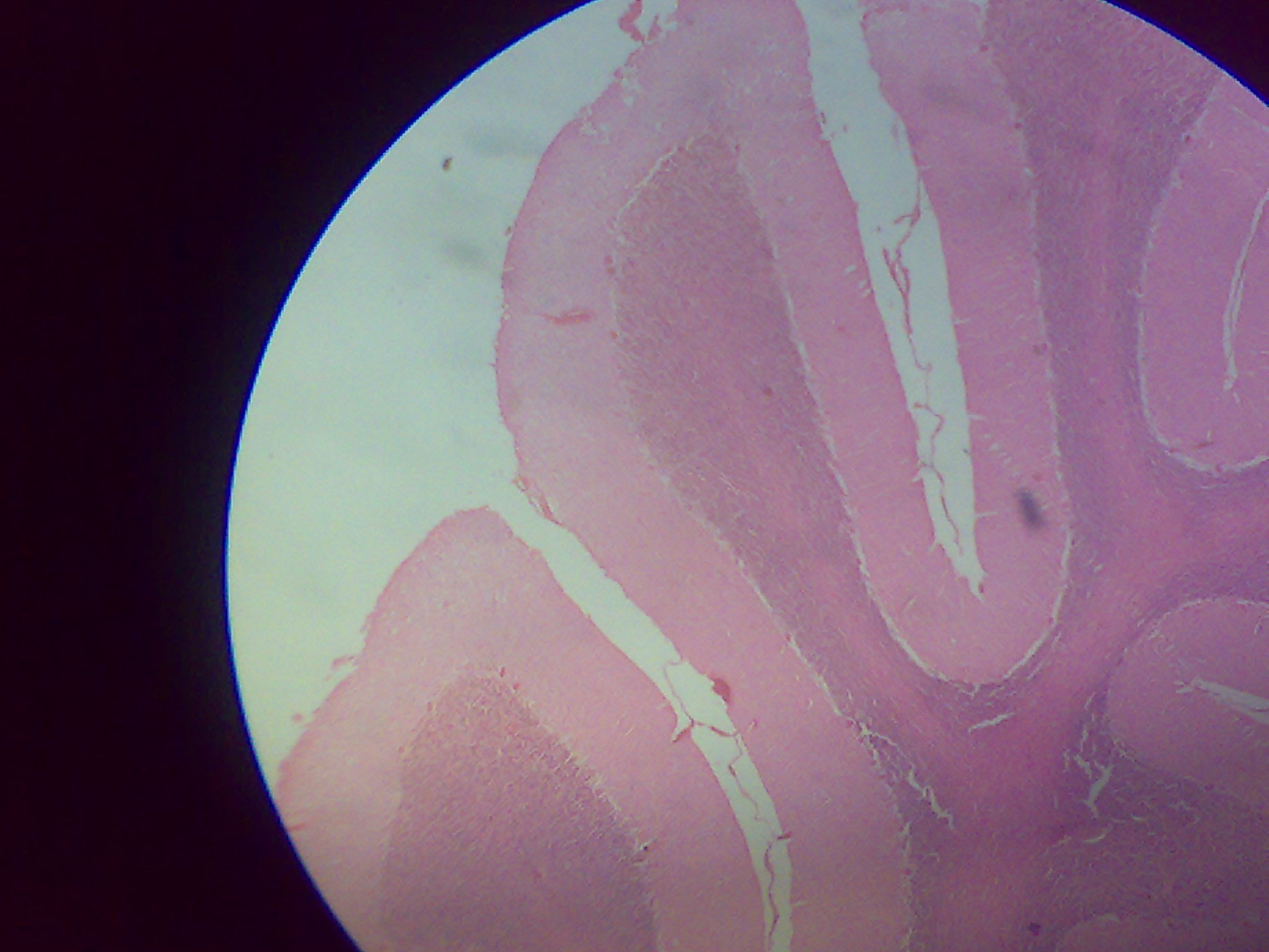
Spinal cord — cross section with gray matter horns
Stain: H&E·Magnification: 4x·Tissue: CNS — gray + white matter·4 labeled regions
Wikimedia Commons · File:Spinal_Marrow.JPG · https://commons.wikimedia.org/wiki/File:Spinal_Marrow.JPG (CC-licensed)
Open the interactive viewer
Sign up free to pan/zoom up to 10×, tap each labeled marker for the structure's description and clinical context, and switch to quiz mode to test yourself on every region.
Create free accountDescription
Central gray matter forms an H/butterfly with anterior (motor) horns wider than posterior (sensory) horns. Surrounded by white matter columns carrying ascending sensory + descending motor tracts.
Labeled regions (4)
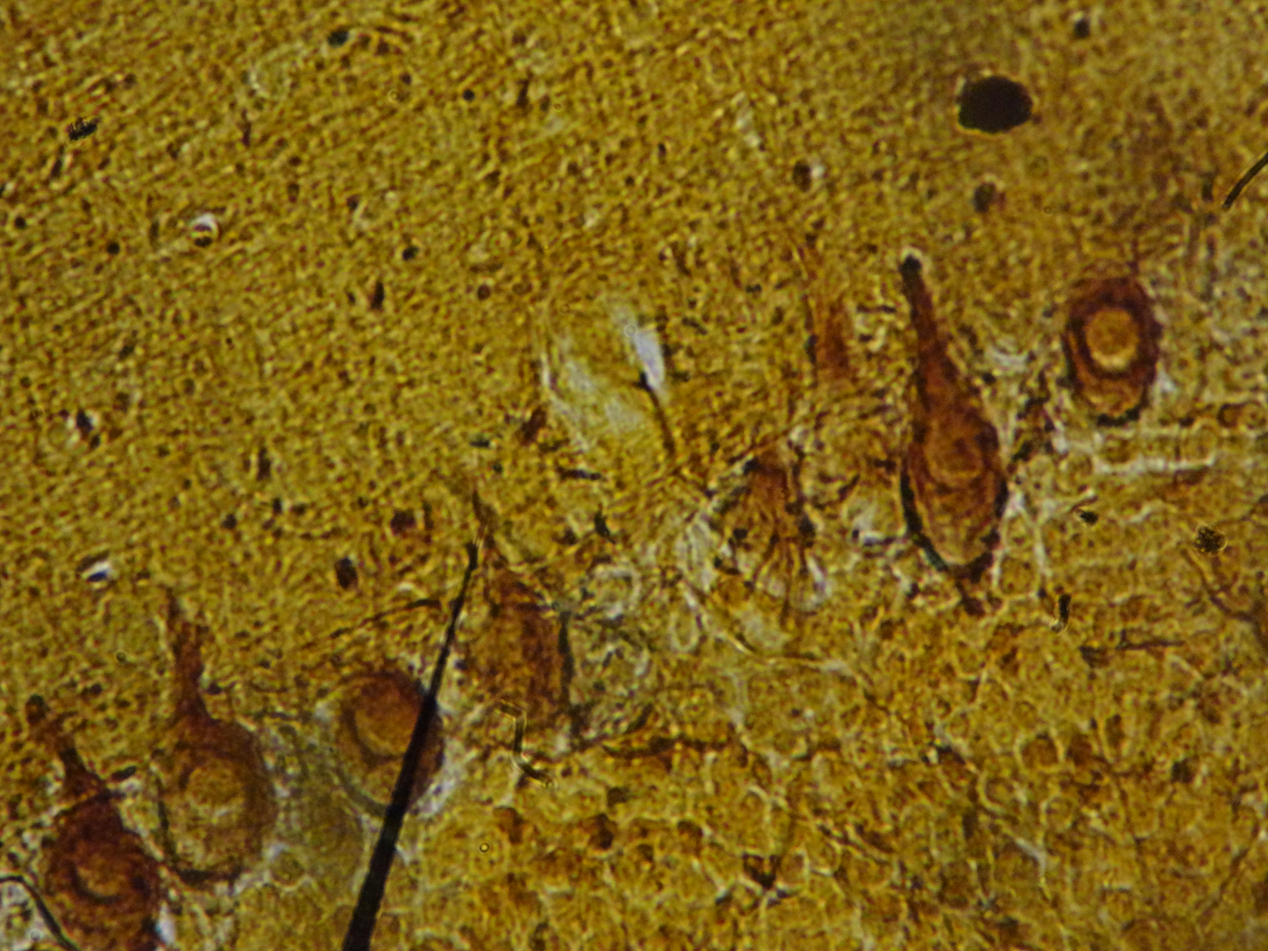
- 1Anterior (ventral) horn
Houses lower motor neuron cell bodies. Destroyed in poliomyelitis + ALS (combined with corticospinal tract loss).
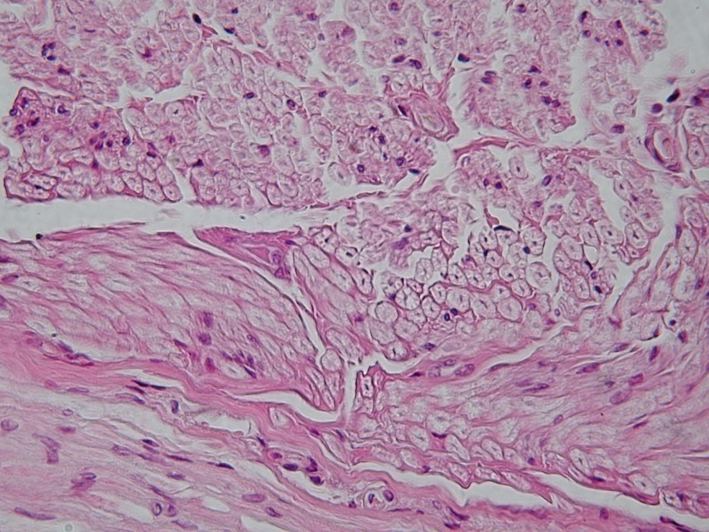
- 2Posterior (dorsal) horn
First-order sensory neurons synapse here. Inputs arrive via dorsal root from DRG.
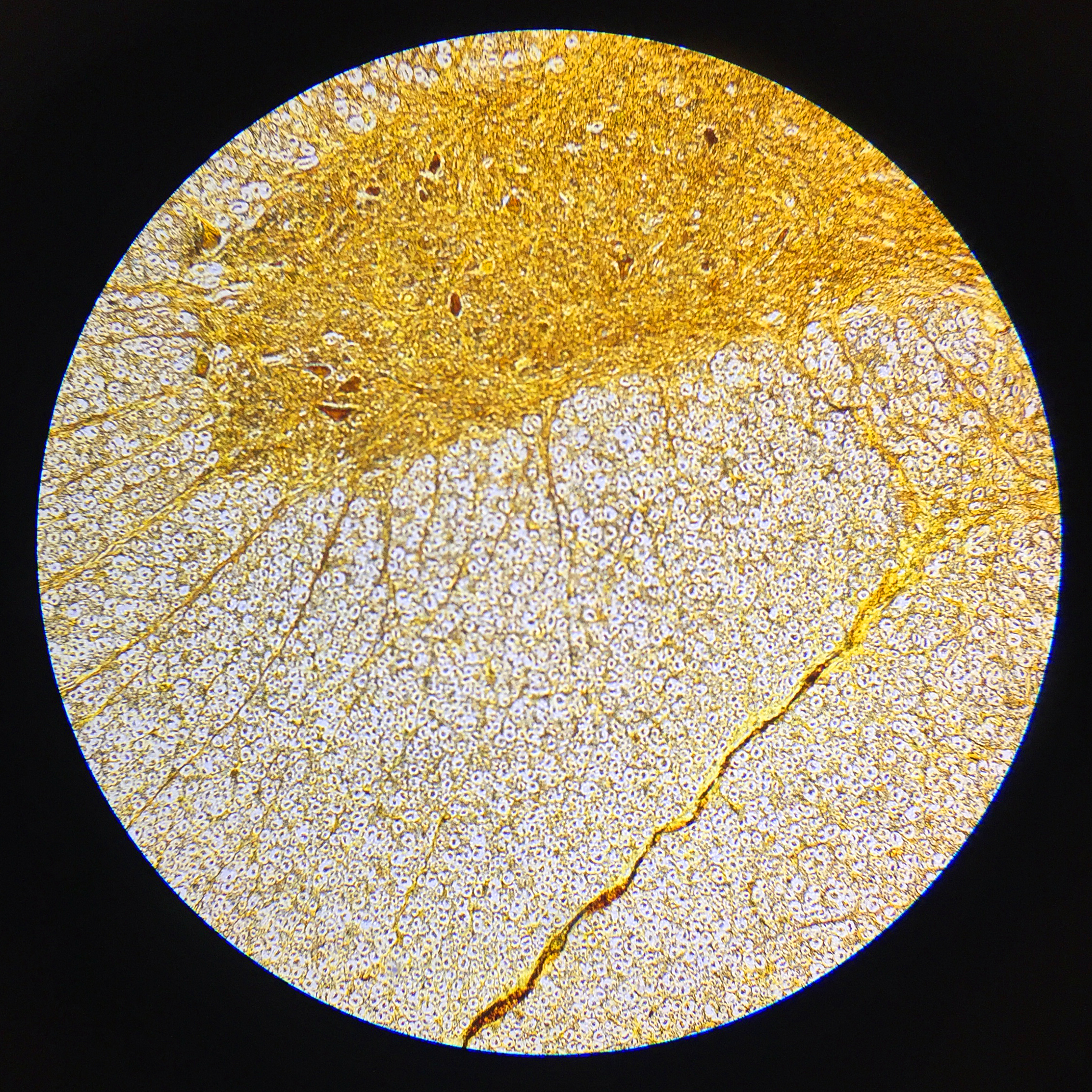
- 3Central canal
Ependyma-lined remnant of the embryonic neural tube; CSF continuous with 4th ventricle.
- 4White matter columns
Pale-staining myelinated tracts. Dorsal columns (fine touch/proprio), lateral corticospinal (UMN), spinothalamic (pain/temp).