Normal·Neurology·Cerebrum
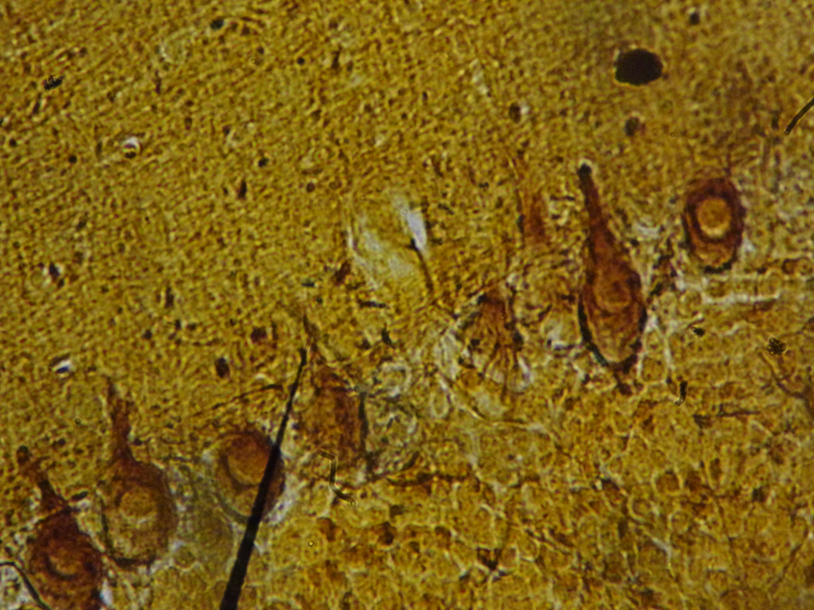
Cerebral cortex — pyramidal neurons
Stain: H&E·Magnification: 40x·Tissue: CNS — six-layered neocortex·3 labeled regions
Wikimedia Commons · File:Pyramidal_cells_-_brain_tissue.jpg · https://commons.wikimedia.org/wiki/File:Pyramidal_cells_-_brain_tissue.jpg (CC-licensed)
Open the interactive viewer
Sign up free to pan/zoom up to 10×, tap each labeled marker for the structure's description and clinical context, and switch to quiz mode to test yourself on every region.
Create free accountDescription
Pyramidal neurons are the principal projection neurons of cerebral cortex. Triangular soma with a single apical dendrite directed toward the pial surface. Large in layer V (Betz cells in motor cortex).
Labeled regions (3)
- 1Pyramidal neuron soma
Triangular cell body with pale nucleus + prominent nucleolus. Glutamatergic projection neuron.
- 2Apical dendrite
Single thick process aimed at pial surface; integrates inputs across cortical layers.
- 3Neuropil
Tangled background of axons, dendrites, glial processes. Lacks discrete cell bodies — looks "fuzzy" pink.