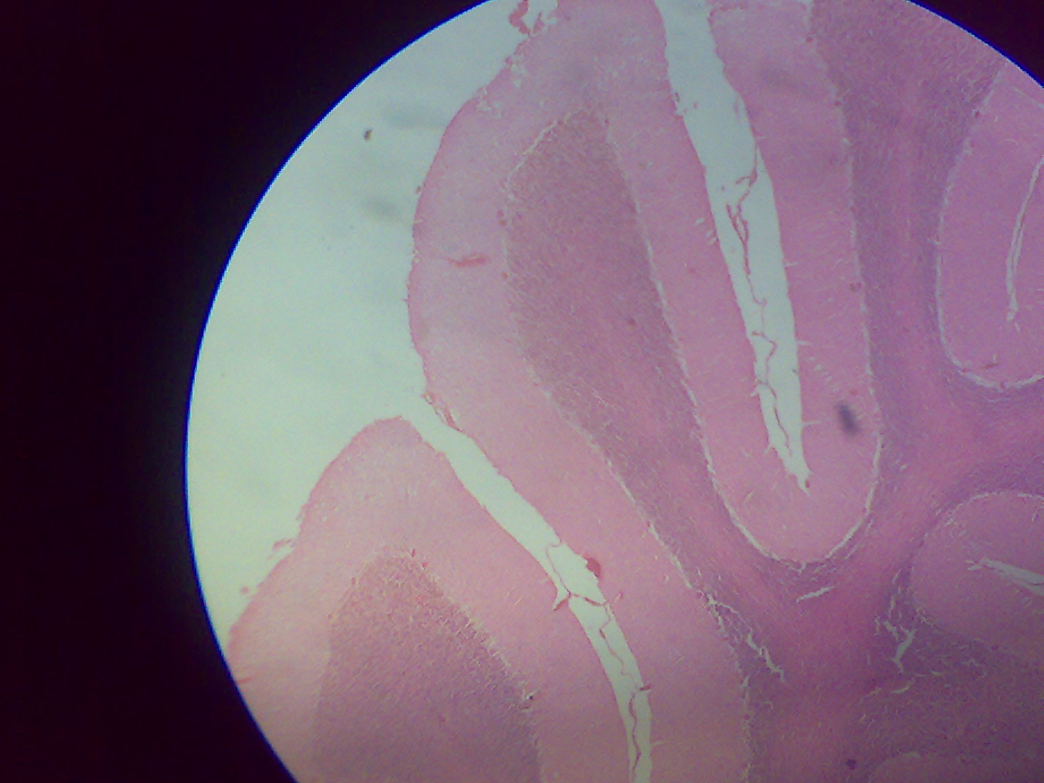
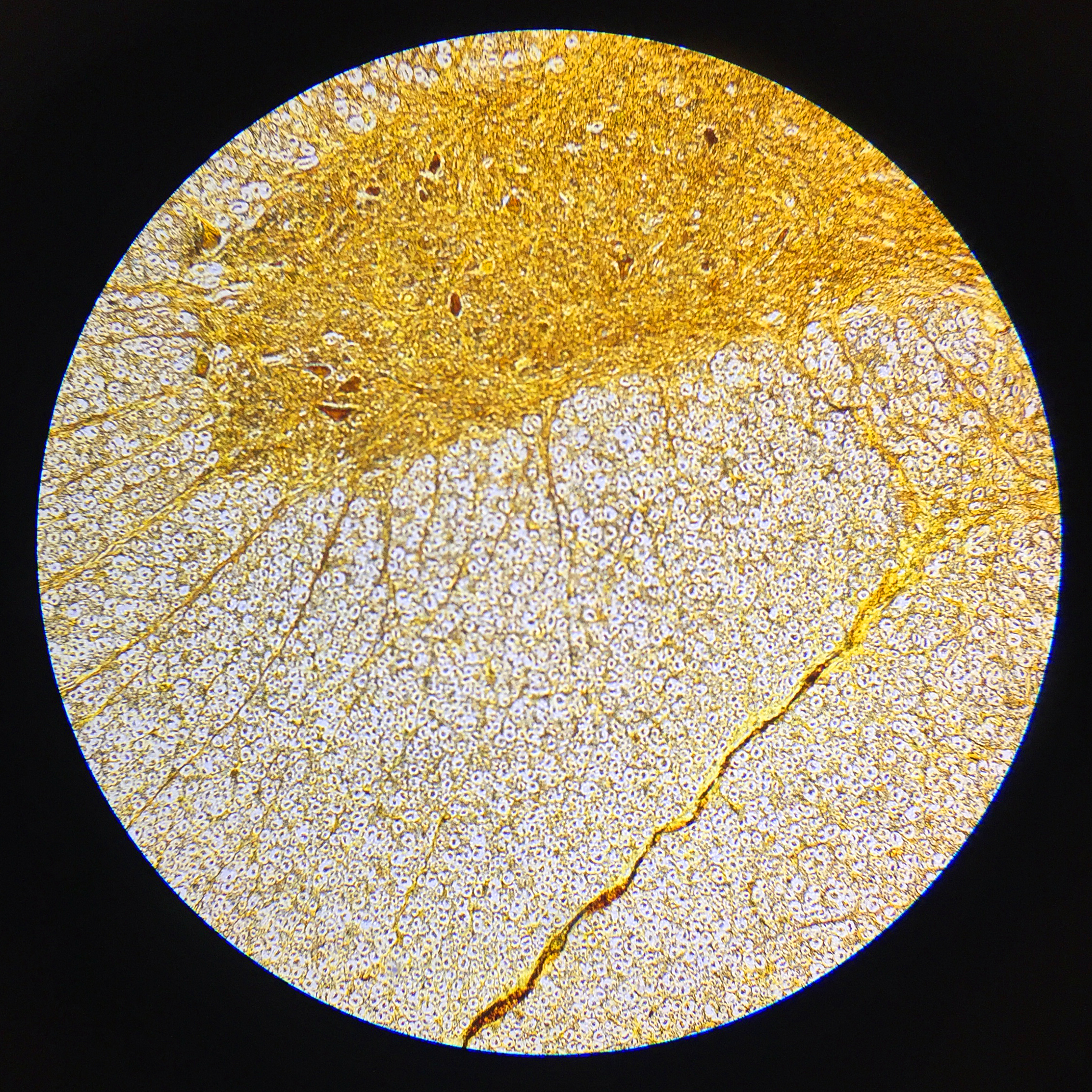
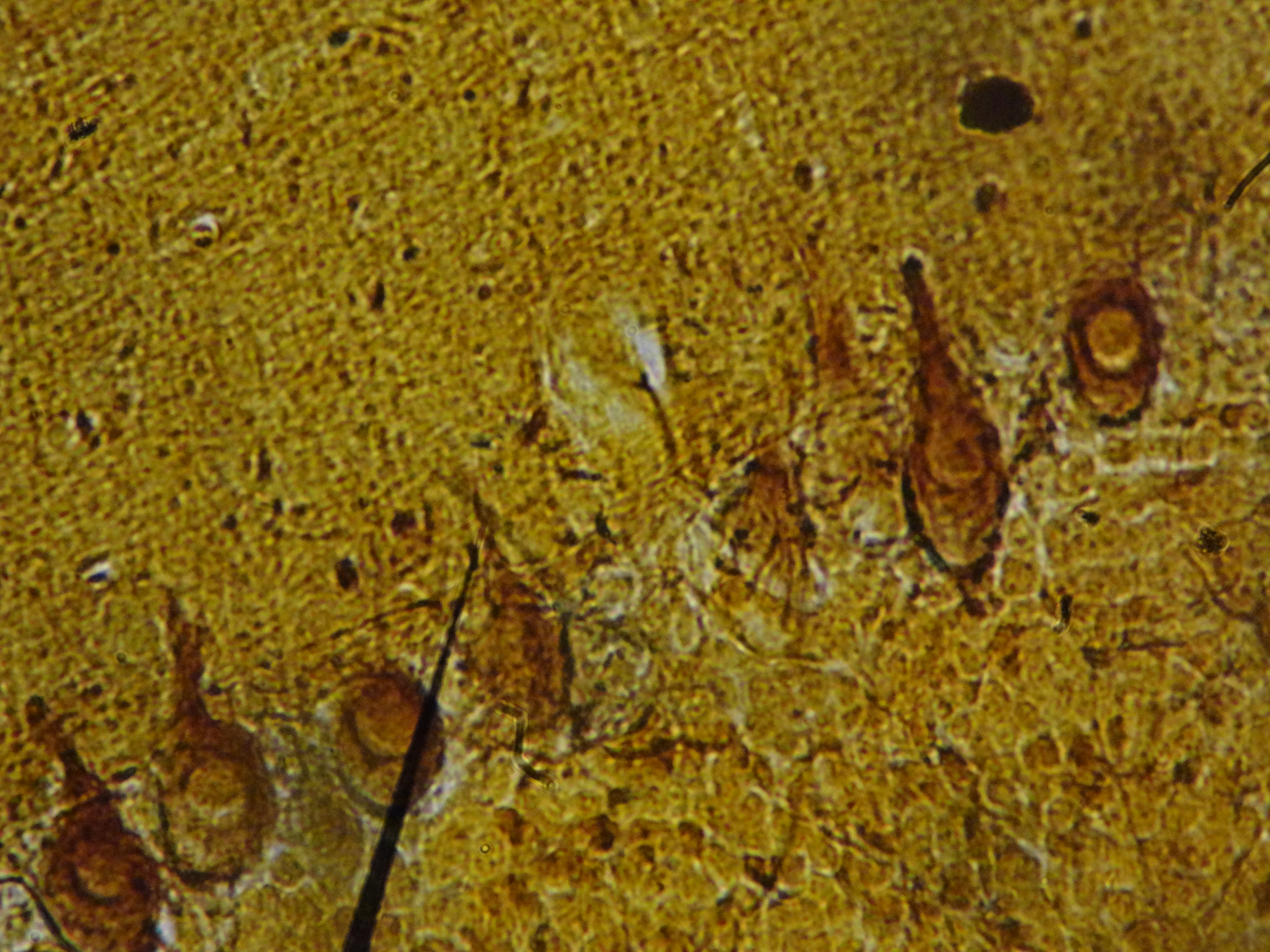
Normal·Neurology·Peripheral nerve
Peripheral nerve — cross section
Stain: H&E·Magnification: 20x·Tissue: Mixed nerve fascicles·4 labeled regions
Wikimedia Commons · File:Peripheral nerve, cross section.jpg · https://commons.wikimedia.org/wiki/File:Peripheral_nerve,_cross_section.jpg (CC-licensed)
Open the interactive viewer
Sign up free to pan/zoom up to 10×, tap each labeled marker for the structure's description and clinical context, and switch to quiz mode to test yourself on every region.
Create free accountDescription
Bundles of myelinated + unmyelinated axons surrounded by three connective tissue sheaths. Each axon is wrapped by a Schwann cell.
Labeled regions (4)
- 1Axon
Central pale dot.
- 2Myelin sheath
Schwann cell plasma membrane wraps the axon (PNS).
- 3Endoneurium
Loose CT around individual axons.
- 4Perineurium
Dense epithelioid sheath around each fascicle; blood–nerve barrier.