Normal·Neurology·Cerebellum
Cerebellum — Purkinje cell layer
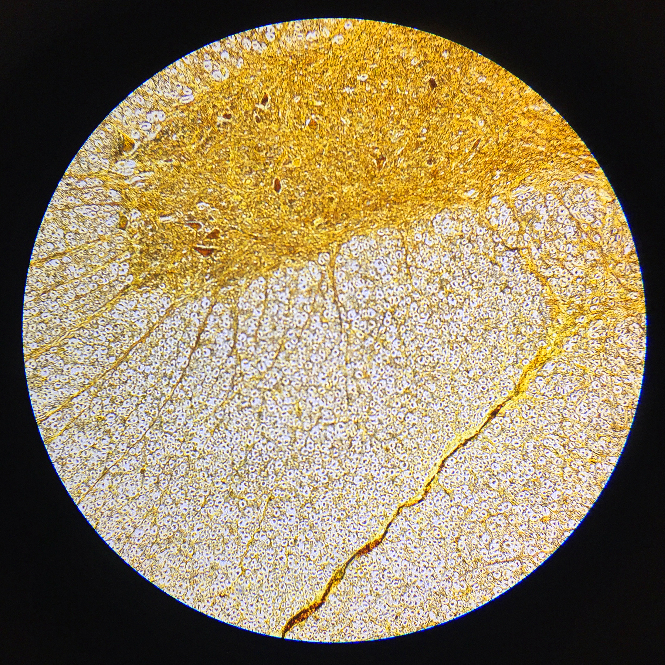
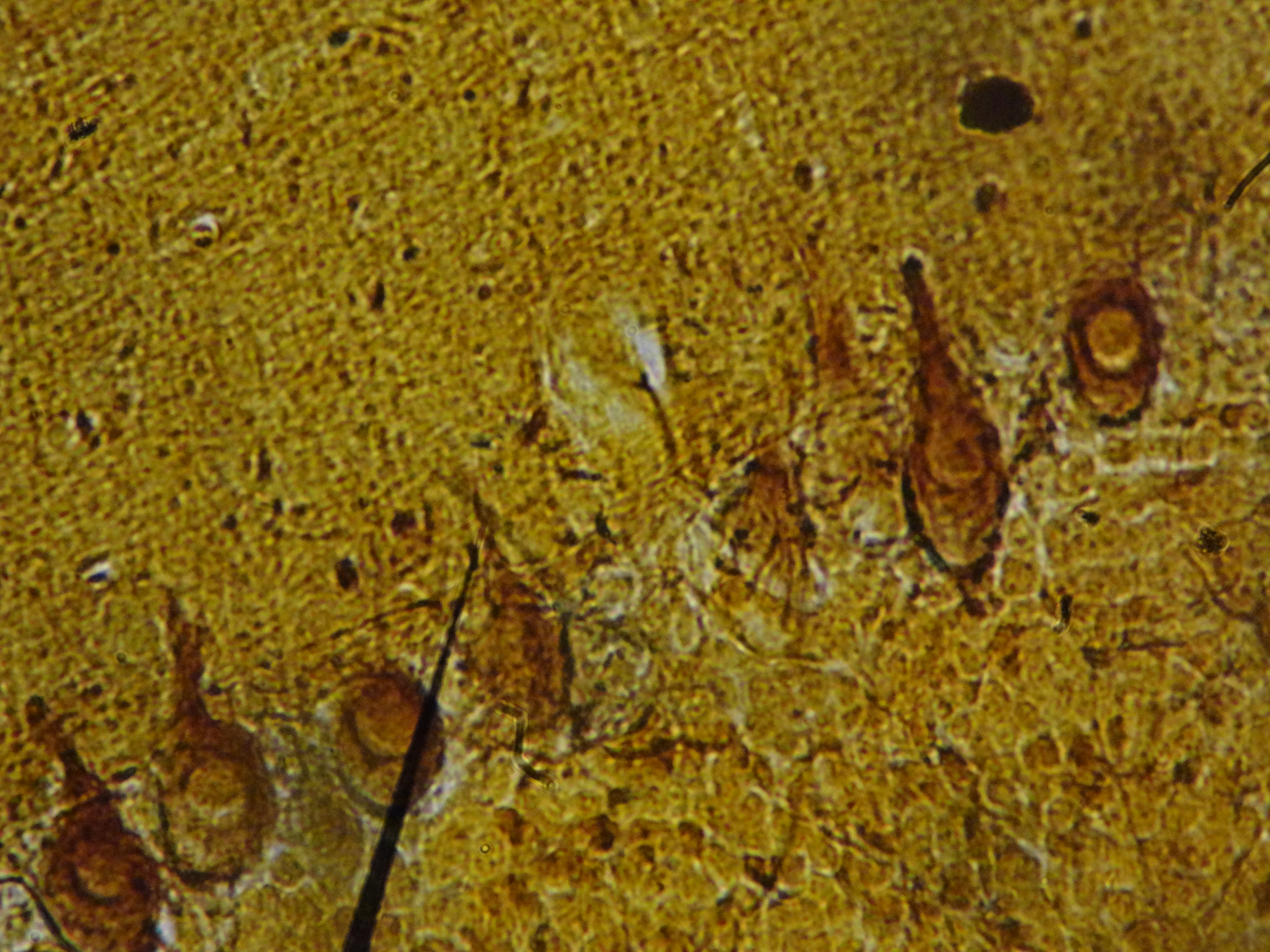
Stain: H&E·Magnification: 20x·Tissue: Cerebellar cortex (molecular, Purkinje, granular layers)·4 labeled regions
Wikimedia Commons · File:Cerebellum_histology_1.jpg · https://commons.wikimedia.org/wiki/File:Cerebellum_histology_1.jpg (CC-licensed)
Open the interactive viewer
Sign up free to pan/zoom up to 10×, tap each labeled marker for the structure's description and clinical context, and switch to quiz mode to test yourself on every region.
Create free accountDescription
Three-layer cerebellar cortex with hallmark Purkinje cell layer wedged between the eosinophilic molecular layer (sparse cells) and densely packed granular layer. Purkinje cells are the only output neurons of the cerebellar cortex.
Labeled regions (4)
- 1Molecular layer
Outermost; sparse neurons + lots of unmyelinated axons (climbing + parallel fibers). Eosinophilic neuropil dominates.
- 2Purkinje cell
Single row of giant flask-shaped neurons; dendrites fan into the molecular layer. Only output of cerebellar cortex (GABAergic). Lost in chronic alcoholism, paraneoplastic anti-Yo, SCA.
- 3Granular layer
Dense small-blue-cell layer of granule neurons (smallest in the body). Their parallel fibers ascend to synapse on Purkinje dendrites.
- 4White matter (medulla)
Myelinated axons entering/leaving the folium. Carries climbing fibers from inferior olive + mossy fibers from pons/spinal cord.