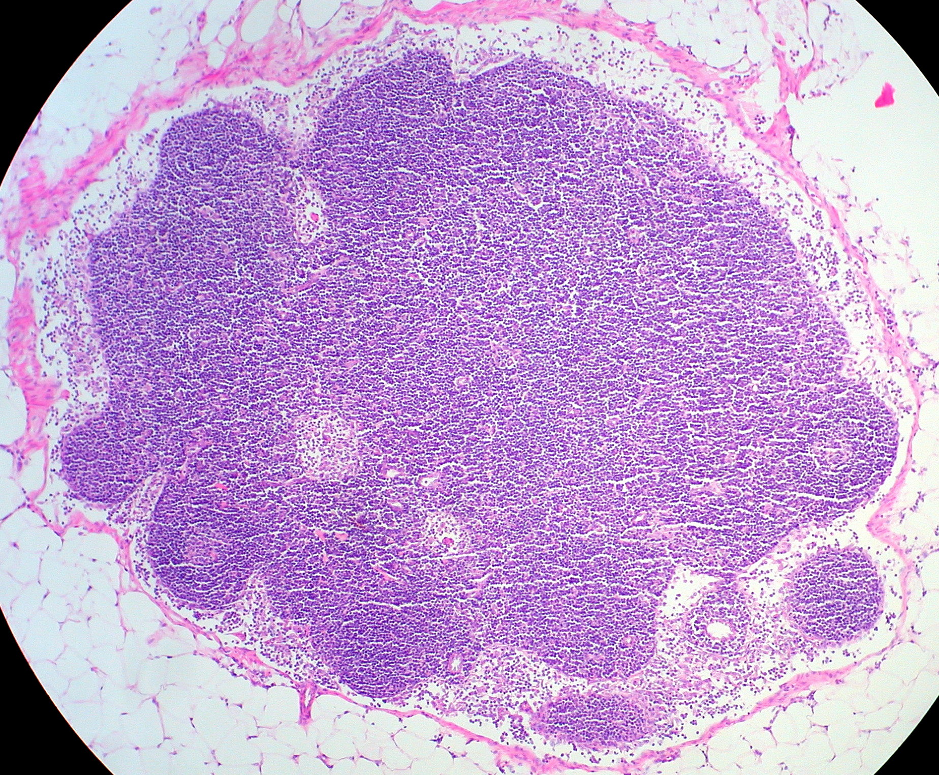
Normal·Hematology/Immunology·Spleen
Spleen — white pulp + red pulp
Stain: H&E·Magnification: 10x·Tissue: Lymphoreticular·3 labeled regions
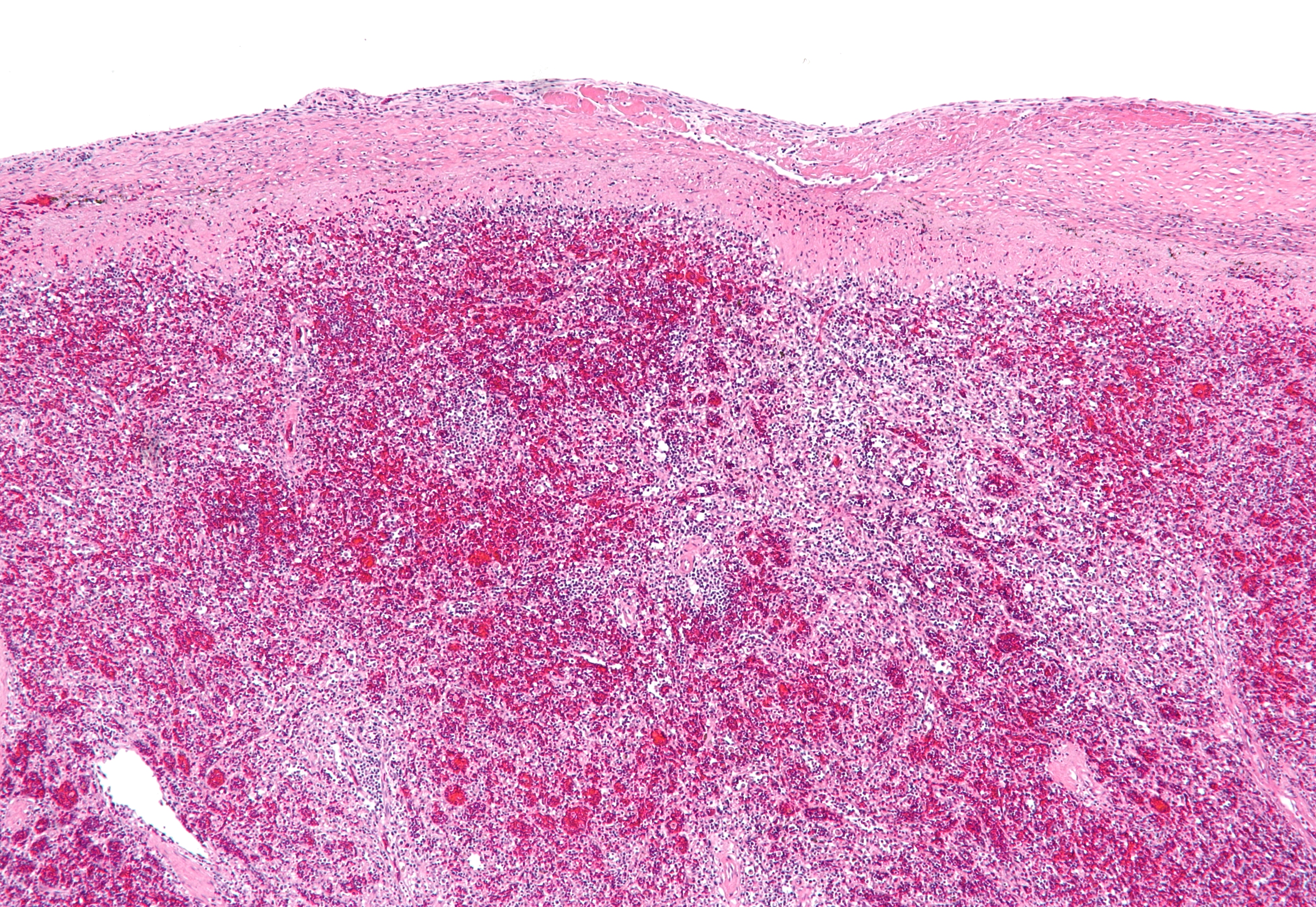
Wikimedia Commons · File:Spleen hyaloserositis - low mag.jpg · https://commons.wikimedia.org/wiki/File:Spleen_hyaloserositis_-_low_mag.jpg (CC-licensed)
Open the interactive viewer
Sign up free to pan/zoom up to 10×, tap each labeled marker for the structure's description and clinical context, and switch to quiz mode to test yourself on every region.
Create free accountDescription
White pulp = PALS (T cells) + B-cell follicles around central arterioles. Red pulp = cords of Billroth + sinusoids filtering blood.
Labeled regions (3)
- 1White pulp (PALS)
Periarteriolar lymphoid sheath — T-cell zone.
- 2Central arteriole
Sits within PALS — diagnostic feature.
- 3Red pulp / sinusoids
Macrophages remove old RBCs; Howell-Jolly bodies appear in asplenia.