Pathologic·Hematology/Immunology·Peripheral blood
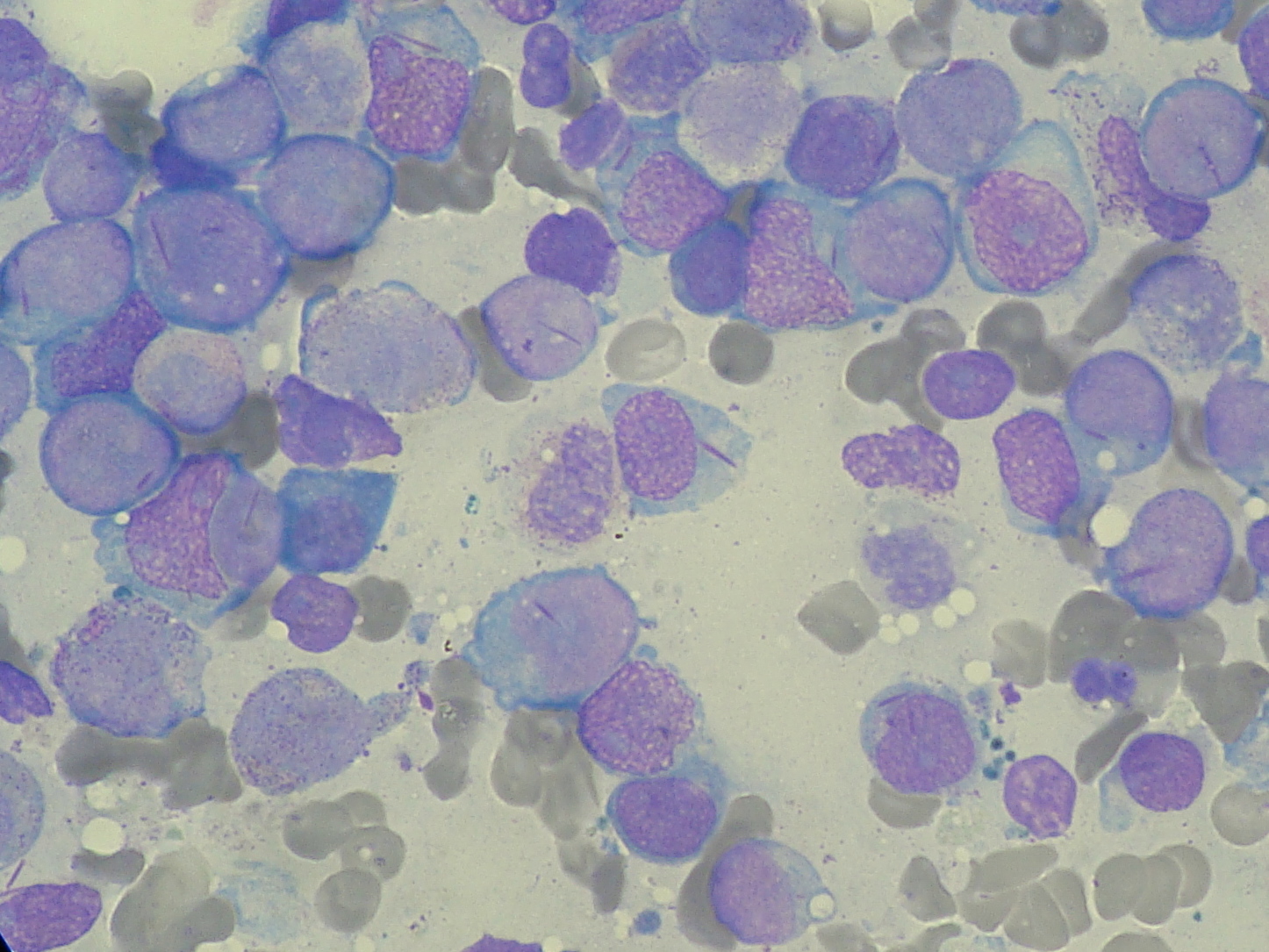
Peripheral smear — AML with Auer rods
Stain: Wright-Giemsa·Magnification: 100x oil·Tissue: Blood smear·3 labeled regions
Wikimedia Commons · File:Myeloblast with Auer rod smear 2010-01-27.JPG · https://commons.wikimedia.org/wiki/File:Myeloblast_with_Auer_rod_smear_2010-01-27.JPG (CC-licensed)
Open the interactive viewer
Sign up free to pan/zoom up to 10×, tap each labeled marker for the structure's description and clinical context, and switch to quiz mode to test yourself on every region.
Create free accountDescription
Sheets of myeloblasts with high nuclear-cytoplasmic ratio, prominent nucleoli, and pink rod-shaped inclusions (Auer rods) — pathognomonic for AML.
Labeled regions (3)
- 1Myeloblast
Large cell, scant cytoplasm, fine chromatin, prominent nucleoli.
- 2Auer rod
Pink azurophilic rod = fused primary granules; pathognomonic for AML (esp. APML M3).
- 3Nucleolus
Prominent — classic for blasts.