Normal·Hematology/Immunology·Lymph node
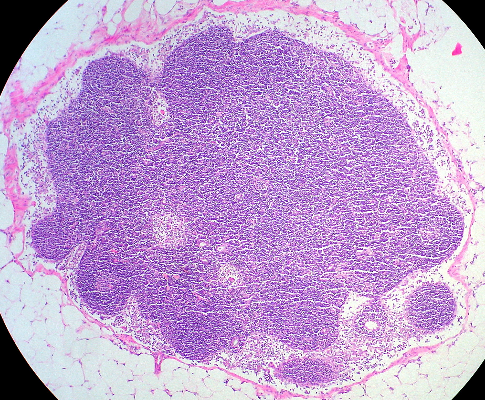
Lymph node — cortex / paracortex / medulla
Stain: H&E·Magnification: 10x·Tissue: Secondary lymphoid·4 labeled regions
Wikimedia Commons · File:Normal Lymph Node.jpg · https://commons.wikimedia.org/wiki/File:Normal_Lymph_Node.jpg (CC-licensed)
Open the interactive viewer
Sign up free to pan/zoom up to 10×, tap each labeled marker for the structure's description and clinical context, and switch to quiz mode to test yourself on every region.
Create free accountDescription
B-cell follicles (cortex) → T-cell paracortex → plasma-cell-rich medulla. Architecture is the basis for differential dx of lymphoma vs reactive hyperplasia.
Labeled regions (4)
- 1Cortex with germinal center
B-cell follicle; active in immune response.
- 2Paracortex
T-cell zone; underdeveloped in DiGeorge.
- 3Medullary cords
Plasma cells + macrophages.
- 4Subcapsular sinus
Receives afferent lymph.