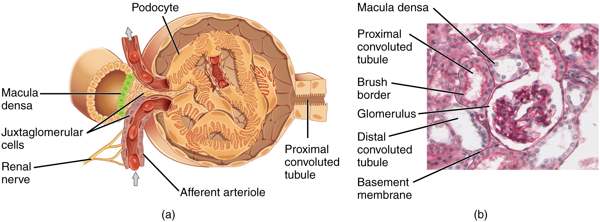
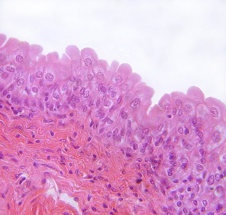
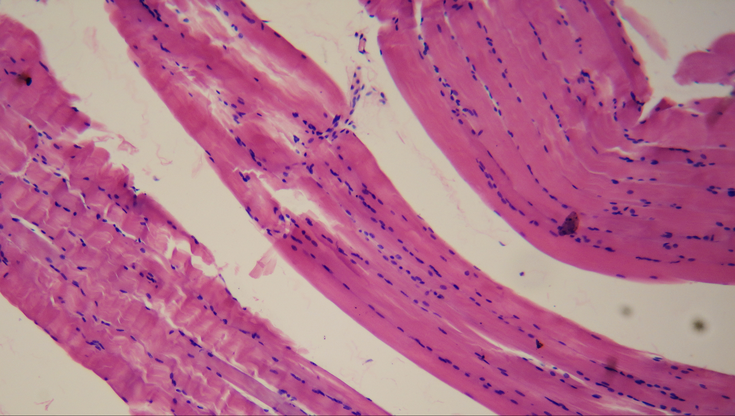
Normal·Renal/Urinary·Bladder
Smooth muscle — bladder wall (detrusor)
Stain: H&E·Magnification: 20x·Tissue: Smooth muscle·3 labeled regions
Wikimedia Commons · File:Smooth muscle (histology slide).jpg by Juan Carlos Fonseca Mata · https://commons.wikimedia.org/wiki/File:Smooth_muscle_(histology_slide).jpg (CC BY-SA 4.0)
Open the interactive viewer
Sign up free to pan/zoom up to 10×, tap each labeled marker for the structure's description and clinical context, and switch to quiz mode to test yourself on every region.
Create free accountDescription
Spindle-shaped cells with single central nuclei and no striations. Involuntary; can undergo hyperplasia (unlike striated muscle which only hypertrophies).
Labeled regions (3)
- 1Spindle-shaped fiber
Tapered ends, single central nucleus; cells overlap so adjacent slices look like 'eyes'.
- 2Central elongated nucleus
Cigar-shaped; contracts into a corkscrew during contraction.
- 3No striations
Distinguishes from cardiac + skeletal — actin/myosin not in register.