Normal·Respiratory·Trachea
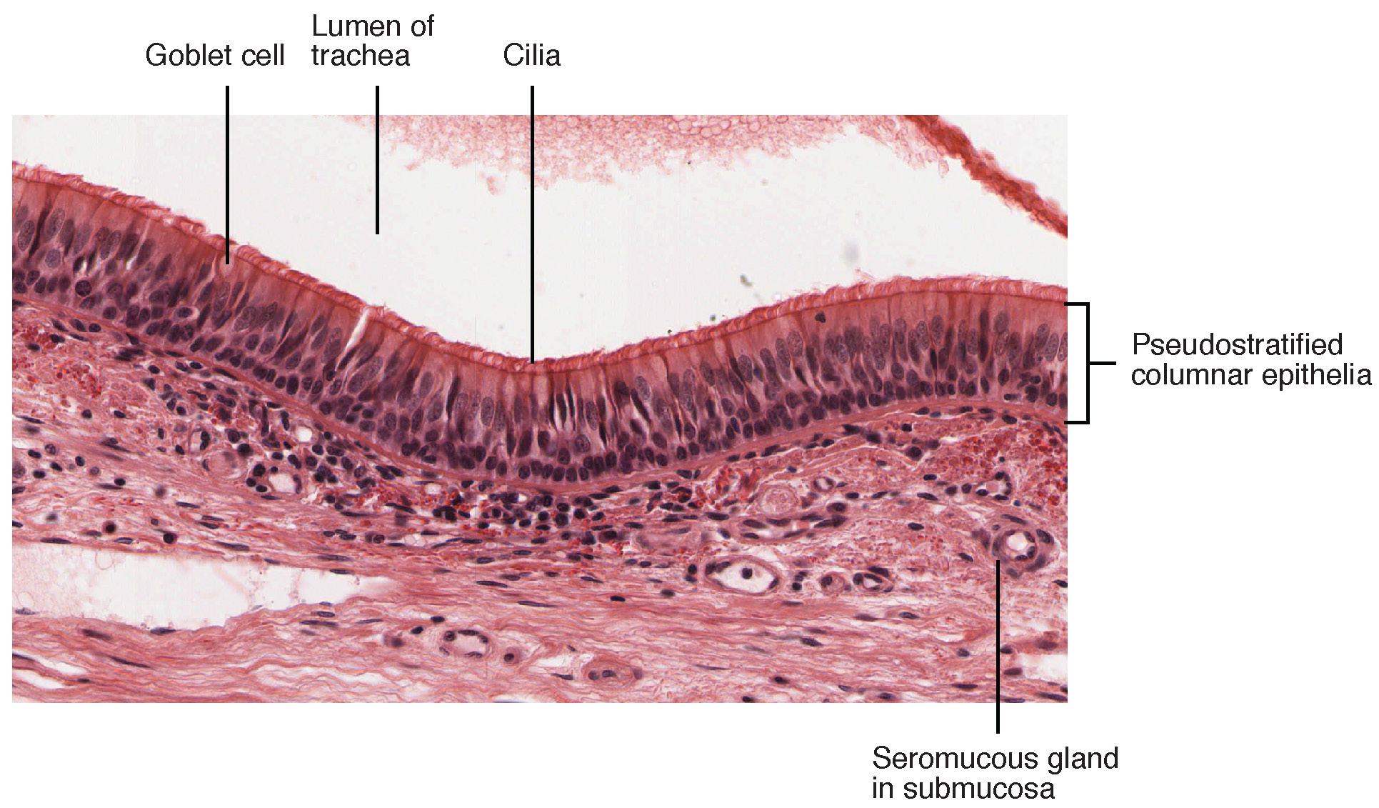
Pseudostratified ciliated columnar epithelium — trachea
Stain: H&E·Magnification: 40x·Tissue: Pseudostratified ciliated columnar·4 labeled regions
Wikimedia Commons · File:2304 Pseudostratified Epithelium.jpg · https://commons.wikimedia.org/wiki/File:2304_Pseudostratified_Epithelium.jpg (CC BY 3.0)
Open the interactive viewer
Sign up free to pan/zoom up to 10×, tap each labeled marker for the structure's description and clinical context, and switch to quiz mode to test yourself on every region.
Create free accountDescription
All cells touch the basement membrane but their nuclei sit at different levels, creating a false sense of stratification. Cilia + goblet cells form the mucociliary escalator.
Labeled regions (4)
- 1Cilia
Motile 9+2 microtubule arrays; beat mucus toward pharynx.
- 2Goblet cell
Mucin secretion; hyperplasia in chronic bronchitis.
- 3Basal cell
Reserve stem cells along basement membrane.
- 4Basement membrane
Thickened in asthma; helps distinguish from chronic bronchitis.