Normal·Respiratory·Lung
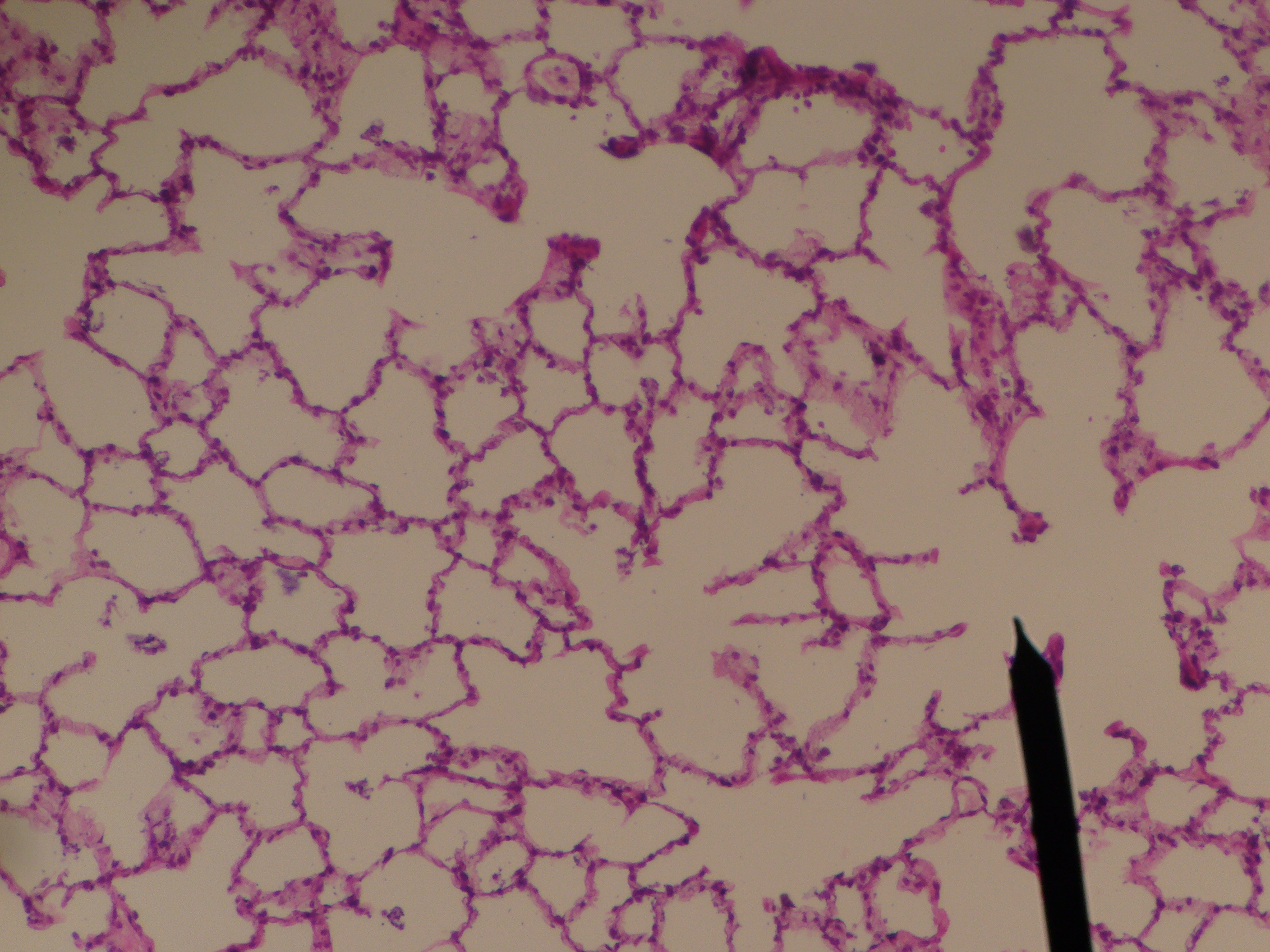
Lung — alveolar parenchyma
Stain: H&E·Magnification: 40x·Tissue: Alveolar·4 labeled regions
Wikimedia Commons · File:Alveolar sac.JPG · https://commons.wikimedia.org/wiki/File:Alveolar_sac.JPG (CC-licensed)
Open the interactive viewer
Sign up free to pan/zoom up to 10×, tap each labeled marker for the structure's description and clinical context, and switch to quiz mode to test yourself on every region.
Create free accountDescription
Open spaces lined by Type I pneumocytes (thin, gas exchange) and Type II (cuboidal, surfactant). Macrophages roam the airspaces.
Labeled regions (4)
- 1Alveolar space
Air-filled lumen.
- 2Type I pneumocyte
Flat squamous cell covering ~97% of alveolar surface.
- 3Type II pneumocyte
Cuboidal; surfactant + progenitor for type I.
- 4Alveolar macrophage
Dust cell; can become 'heart failure cells' (hemosiderin-laden) in chronic LHF.