Pathologic·Respiratory·Bronchus
Asthma — airway remodeling + eosinophilic inflammation
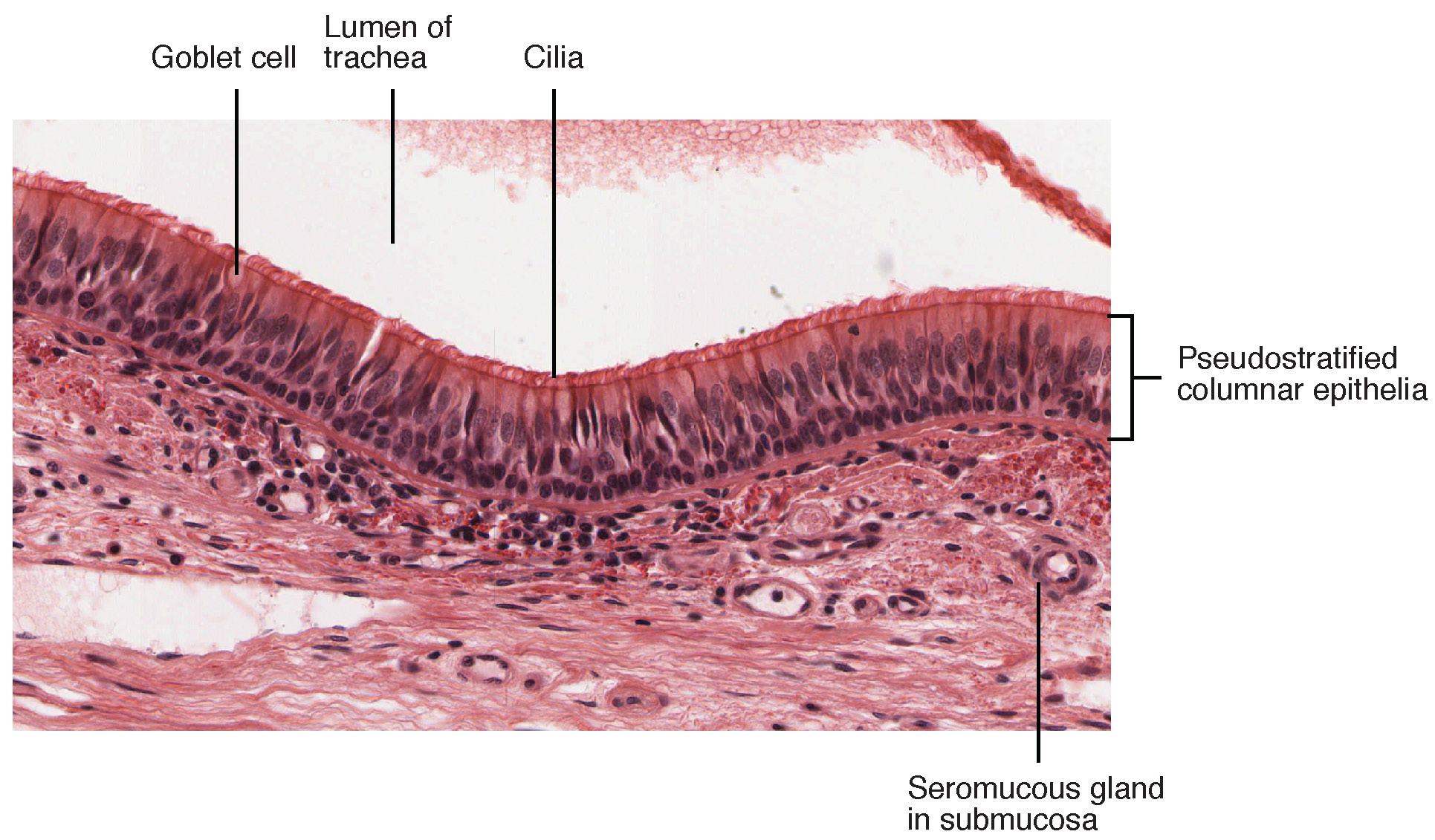
Stain: H&E·Magnification: 20x·Tissue: Bronchial wall with goblet cell metaplasia·4 labeled regions
Wikimedia Commons · File:Asthma.jpg · https://commons.wikimedia.org/wiki/File:Asthma.jpg (CC-licensed)
Open the interactive viewer
Sign up free to pan/zoom up to 10×, tap each labeled marker for the structure's description and clinical context, and switch to quiz mode to test yourself on every region.
Create free accountDescription
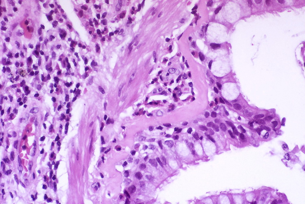
Chronic Th2-driven airway disease: goblet cell metaplasia + mucus plugging, basement membrane thickening, smooth muscle hyperplasia, and eosinophilic infiltrate (with Charcot-Leyden crystals / Curschmann spirals in sputum).
Labeled regions (4)
- 1Goblet cell metaplasia
Expanded mucus-secreting cell population in surface epithelium. Mucus plugging contributes to obstruction.
- 2Thickened basement membrane
Subepithelial fibrosis (collagen I/III/V deposition). Hallmark of chronic airway remodeling.
- 3Smooth muscle hyperplasia
Thickened muscle band drives bronchoconstriction. Targeted by SABA / LABA β2 agonists.
- 4Eosinophilic infiltrate
Bright pink granular cells in submucosa. IL-5 driven; targeted by mepolizumab / benralizumab.