Viral & Autoimmune Hepatitis
A high-yield Step 1/2 CK walkthrough of viral hepatitis (A–E) and autoimmune hepatitis, moving from pathophysiology to serology/autoantibody interpretation, classic vignettes, and next-best-step management. Emphasizes HBV serology (including the window period), HCV RNA confirmation before pangenotypic DAA therapy, and AIH diagnosis and steroid therapy.
Two Roads to Hepatocyte Injury
Hepatitis is inflammation of hepatocytes, signaled by a hepatocellular enzyme pattern (ALT/AST up, ALP relatively spared). Boards split the causes into infectious (five hepatotropic viruses, A–E) and immune-mediated (autoimmune hepatitis, AIH). The first fork is tempo: acute, self-limited injury (HAV, HEV, most adult HBV) versus chronic disease that smolders toward fibrosis → cirrhosis → hepatocellular carcinoma (HCC) (HBV, HCV, HDV, untreated AIH).
Acute histology is shared: ballooning degeneration, spotty lobular necrosis, and Councilman (apoptotic/acidophil) bodies; chronic disease adds a portal lymphocytic infiltrate and bridging fibrosis. Jaundice, dark urine (conjugated bilirubinuria), and RUQ discomfort are common to all. So recognize the pattern first — then let epidemiology (travel, IVDU, sex, pregnancy), serology, and autoantibodies name the cause and drive the next-best step.
- HAV — picornavirus, naked +ssRNA; fecal–oral (shellfish, travel, daycare). Acute only, never chronic. Anti-HAV IgM = active, IgG = immunity. Inactivated (killed) vaccine.
- HBV — hepadnavirus, partial dsDNA, replicates via reverse transcriptase; blood/sexual/perinatal. Chronicity rises as host age falls: ~90% of neonates vs <5% of adults. Integrates into host genome → HCC even without cirrhosis. Extrahepatic: PAN, membranous nephropathy.
- HCV — flavivirus, +ssRNA; IVDU is #1. Most become chronic (~75–85%). Extrahepatic: mixed cryoglobulinemia, MPGN, porphyria cutanea tarda, lichen planus. DAAs cure >95%.
- HDV — defective circular −ssRNA; requires HBsAg. Coinfection (with acute HBV, usually resolves) vs superinfection on chronic HBV (→ severe/fulminant).
- HEV — hepevirus, naked +ssRNA; fecal–oral/waterborne, undercooked pork/game. Acute (chronic if immunosuppressed); fulminant in pregnancy, mortality up to ~20%.
Hepatitis A–E at a Glance
| Virus | Genome / family | Transmission | Chronic? | Board pearl |
|---|---|---|---|---|
| A | +ssRNA, picornavirus | Fecal–oral | No | Traveler/shellfish; self-limited; IgM = acute |
| B | partial dsDNA, hepadnavirus | Blood, sex, perinatal | Yes (↑ if young) | HCC without cirrhosis; ground-glass hepatocytes; PAN, membranous nephropathy |
| C | +ssRNA, flavivirus | Blood (IVDU) | Yes (~75–85%) | Cryoglobulinemia, MPGN, PCT; confirm with RNA; DAA cure |
| D | −ssRNA, defective (delta) | Blood/sex (needs HBV) | Only with HBV | Superinfection → fulminant |
| E | +ssRNA, hepevirus | Fecal–oral/water | No* | Fulminant in pregnancy |
- HBsAg — surface antigen → current infection; persistence >6 months = chronic.
- Anti-HBs — immunity (recovery or vaccination).
- HBeAg — active replication / high infectivity; anti-HBe = lower infectivity.
- HBcAg — not detectable in serum. Anti-HBc IgM = acute/recent; anti-HBc IgG = past or chronic exposure.
- Window period — HBsAg has cleared but anti-HBs is not yet positive → anti-HBc IgM is the ONLY positive marker.
- Vaccinated = isolated anti-HBs (no anti-HBc, since the vaccine is surface antigen only).
- HBV DNA quantifies viral load → guides treatment and monitoring.
Shortcut: anti-HBc positive + HBsAg negative + anti-HBs negative = think window period.

A. A 30-year-old has malaise, nausea, and jaundice; ALT 1,400. Labs: HBsAg negative, anti-HBs negative, anti-HBc IgM positive, HBeAg negative.
- Dx: acute HBV in the window period.
- Next step: supportive care and repeat serology — most immunocompetent adults clear the virus (anti-HBs seroconversion). Antivirals only for fulminant/severe acute HBV.
B. A 45-year-old former IV drug user is found to be anti-HCV positive on routine screening; LFTs near-normal.
- Next best step: HCV RNA (PCR) to confirm active infection — anti-HCV cannot distinguish active from cleared disease.
- If RNA-positive → assess fibrosis (e.g., FIB-4 or elastography) and treat with pangenotypic direct-acting antivirals (DAAs); routine genotyping is no longer required for most regimens. Start HCC surveillance if cirrhosis is present.
Autoimmune Hepatitis (AIH)
AIH is a T-cell–mediated attack on hepatocytes, classically in women (bimodal: adolescence and middle age) and frequently associated with other autoimmune disease (Hashimoto thyroiditis, celiac, type 1 DM, RA). Presentation spans asymptomatic transaminitis → fatigue/arthralgia → acute fulminant hepatitis.
Labs: hepatocellular pattern (↑ALT/AST, ALP relatively spared), polyclonal hypergammaglobulinemia with a markedly elevated IgG, and autoantibodies. Type 1 (most common; adults): ANA and/or anti–smooth-muscle (anti-actin) antibody. Type 2 (children; more aggressive): anti–LKM-1 and/or anti–liver cytosol-1.
Diagnosis integrates autoantibodies, high IgG, exclusion of viral hepatitis, and liver biopsy — interface hepatitis with a lymphoplasmacytic infiltrate and hepatocyte rosettes. Untreated disease progresses to cirrhosis.
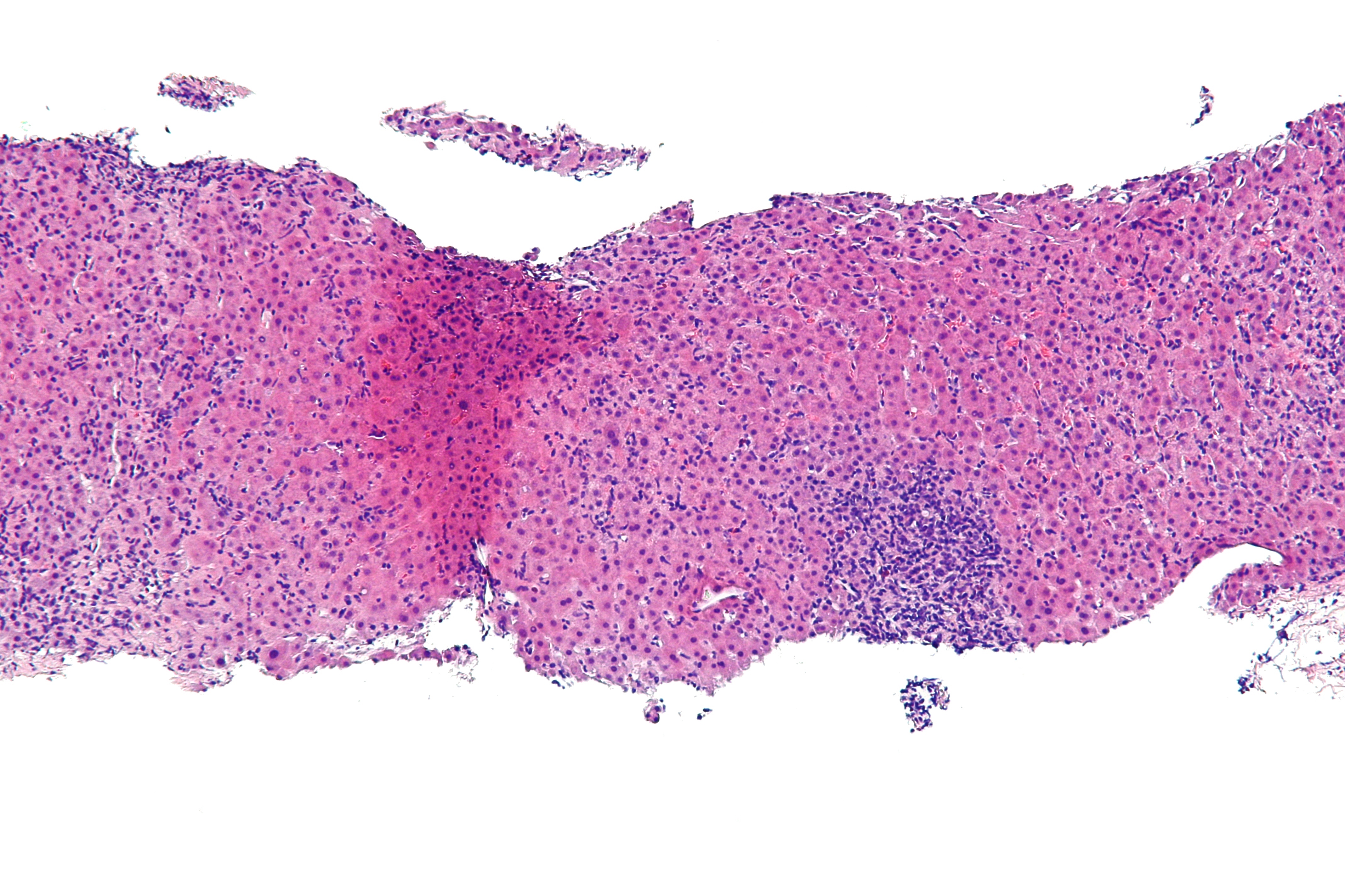
A 28-year-old woman has 2 months of fatigue and arthralgias with mild scleral icterus. AST 620, ALT 780, ALP mildly elevated, IgG markedly high. ANA and anti–smooth-muscle antibody positive; anti-HAV IgM, HBsAg, and anti-HCV all negative.
- Dx: type 1 autoimmune hepatitis.
- Next best step: liver biopsy to confirm (interface hepatitis) and stage fibrosis.
- Treatment: corticosteroids (prednisone) ± azathioprine to induce and maintain remission; budesonide is an option in non-cirrhotics (avoid in cirrhosis due to portosystemic shunting). Titrate by transaminases and IgG. Liver transplant for decompensated/refractory disease (AIH can recur in the graft).
Trap: don’t anchor on “hepatitis” = viral — negative viral serologies + autoantibodies + high IgG in a young woman = AIH.
AIH Type 1 vs Type 2
| Feature | Type 1 | Type 2 |
|---|---|---|
| Typical patient | Adults, women | Children / teens |
| Autoantibodies | ANA, anti–smooth muscle (anti-actin) | Anti–LKM-1, anti–liver cytosol-1 |
| Frequency (Western) | More common | Less common |
| Course | Variable | Often more aggressive |
| Shared | ↑ IgG, interface hepatitis on biopsy, steroid-responsive | same |
- “The vowels hit your bowels.” Hepatitis A and E (the vowels) spread fecal–oral/enteric and are acute, self-limited — no chronic carrier state in immunocompetent hosts.
- Consonants → chronic. B, C, D are blood/body-fluid borne and can turn chronic → cirrhosis / HCC.
- HEV + pregnancy = disproportionately high mortality (up to ~20%).
Management & Prophylaxis — Next-Best Steps
- Acute HAV / HEV: supportive — they self-resolve. Post-exposure HAV prophylaxis: HepA vaccine preferred; add immunoglobulin for infants <12 months, immunocompromised, or chronic liver disease.
- Chronic HBV: treat eligible patients with entecavir or tenofovir (or peg-IFN); goal is HBV DNA suppression. Perinatal prevention: infants of HBsAg-positive mothers get HBV vaccine + HBIG at birth. Needlestick (unvaccinated, HBsAg-positive source): HBIG + vaccine series.
- Chronic HCV: DAAs cure >95%; screen all adults at least once.
- HCC surveillance: liver ultrasound ± AFP every 6 months in cirrhosis and in high-risk chronic HBV.
- AIH: prednisone ± azathioprine for induction/maintenance; taper guided by transaminases and IgG; transplant for end-stage disease.
Bottom line: serology names the virus, autoantibodies + high IgG name AIH — and each diagnosis carries a specific, testable next step.
Practice GI now
Board-style questions, spaced-repetition flashcards, and a Socratic AI tutor — free to start.