Thyroid Nodules & Thyroid Cancer
A Step 2 CK high-yield lesson on thyroid nodules and thyroid cancer, walking from pathophysiology through the TSH-first diagnostic algorithm (uptake scan for low TSH, FNA/Bethesda otherwise) to management, with the four cancer histologies, MEN2, and board buzzwords. Emphasizes next-best-step traps: hot nodules skip FNA, and pheochromocytoma must be excluded before medullary/MEN2 surgery.
Overview & Pathophysiology
Thyroid nodules are extremely common — palpable in ~5% of adults but found incidentally in up to half of all neck imaging studies. The overwhelming majority are benign colloid nodules or follicular adenomas; only ~7–15% are malignant. Autonomously functioning ("hot") nodules make thyroid hormone independent of TSH, suppressing the rest of the gland — and are almost never cancerous.
Malignancy arises from two lineages: follicular epithelium (papillary, follicular, and dedifferentiated anaplastic carcinoma) and parafollicular C cells (medullary carcinoma). Well-differentiated cancers retain two follicular-cell functions that drive management — iodine uptake (enabling radioactive iodine therapy) and thyroglobulin secretion (a post-thyroidectomy tumor marker).
The exam rewards a rigid workup order: characterize the nodule biochemically (TSH) and sonographically before reaching for a needle.
Red flags (raise malignancy suspicion):
- History of head/neck radiation, especially in childhood (or whole-body, e.g., Chernobyl)
- Rapid growth, firm/fixed nodule, or new hoarseness (recurrent laryngeal nerve invasion)
- Cervical lymphadenopathy, dysphagia, or stridor
- Age <20 or >70, male sex, family history of thyroid cancer or MEN2
Reassuring: soft, mobile, part of a multinodular goiter, or a toxic (hot) nodule.
Suspicious ultrasound features:
- Marked hypoechogenicity
- Microcalcifications (correlate with psammoma bodies)
- Irregular/infiltrative margins
- Taller-than-wide shape (AP > transverse)
- Extrathyroidal extension or pathologic nodes
Cystic/spongiform nodules are almost always benign.
TSH-Directed Workup (labs → next step)
| Setting | TSH | Next best step | Interpretation |
|---|---|---|---|
| New nodule, initial | — | TSH + ultrasound (never FNA first) | Directs the entire workup |
| Nodule + low TSH | ↓ | I-123 / Tc-99m uptake scan | Hot → autonomous, ~benign, treat hyperthyroidism; cold → FNA |
| Nodule + normal/high TSH | →/↑ | US-guided FNA per risk pattern + size | Cytology → Bethesda category |
| Suspected medullary | → | Calcitonin + CEA, RET, plasma/urine metanephrines | Exclude pheochromocytoma before surgery |
| Post-thyroidectomy (differentiated) | suppressed (goal) | Thyroglobulin + anti-Tg antibody | Rising Tg = recurrence |
FNA size thresholds (sonographic risk pattern sets the cutoff):
- High/intermediate-suspicion pattern → FNA at ≥1 cm
- Low-suspicion → ≥1.5 cm; very-low/spongiform → ≥2 cm or observe
- Purely cystic nodule → no FNA
Bethesda cytology → action:
- I Nondiagnostic → repeat US-guided FNA
- II Benign → clinical + US surveillance
- III AUS/FLUS → repeat FNA or molecular testing
- IV Follicular neoplasm → molecular testing or diagnostic lobectomy
- V Suspicious for malignancy → surgery
- VI Malignant → surgery
Key trap: follicular carcinoma is diagnosed only by capsular/vascular invasion on the surgical specimen — cytology cannot distinguish it from a benign follicular adenoma.
Stem: A 34-year-old woman has a 1.5-cm firm right-thyroid nodule and a palpable ipsilateral cervical node. TSH is normal. Ultrasound shows a hypoechoic nodule with microcalcifications and a taller-than-wide shape.
Diagnosis: Features point to papillary thyroid carcinoma (lymphatic spread → the node).
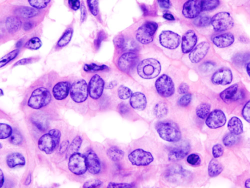
Next best step: US-guided FNA of the nodule (and the abnormal node). Do not order a radioiodine scan — that is only for low TSH. FNA shows Orphan-Annie-eye nuclei, nuclear grooves, and psammoma bodies → Bethesda VI.
Management: thyroidectomy (lobectomy for small low-risk tumors; total thyroidectomy here for nodal disease) with therapeutic dissection of the involved neck compartment ± radioactive iodine ablation, then TSH-suppressive levothyroxine and thyroglobulin surveillance. Prognosis is excellent even with nodal metastases.
Thyroid Cancer Comparison
| Cancer | Cell / spread | Buzzwords | Marker | Notes |
|---|---|---|---|---|
| Papillary (~80%) | Follicular; lymphatic | Orphan-Annie nuclei, psammoma bodies, nuclear grooves; BRAF V600E, RET/PTC; prior radiation | Thyroglobulin | Best prognosis; nodes common but low-risk |
| Follicular (~10%) | Follicular; hematogenous (bone, lung) | Capsular/vascular invasion; RAS, PAX8-PPARγ | Thyroglobulin | FNA can't diagnose → lobectomy |
| Medullary (~5%) | Parafollicular C cells | Amyloid stroma (from calcitonin); RET, MEN2 | Calcitonin, CEA | Not iodine-avid; screen for pheo |
| Anaplastic (~2%) | Undifferentiated follicular | Elderly, rock-hard rapidly enlarging mass; local invasion | None | Dismal prognosis; airway threat |
Stem: A 28-year-old man with episodic headache, palpitations, and sweating is found to have a thyroid nodule; FNA shows sheets of cells in an amyloid stroma. Serum calcitonin and CEA are elevated; his father had a thyroidectomy.
Diagnosis: Medullary thyroid carcinoma in MEN2 (germline RET mutation).
Next best step: Before any thyroid surgery, exclude pheochromocytoma (plasma-free or 24-h urine metanephrines) — operating on an unrecognized pheo can trigger a fatal hypertensive crisis. If a pheo is present, resect it first (after alpha- then beta-blockade).
Management: total thyroidectomy + central neck dissection (MTC is multicentric and not iodine-responsive); check for parathyroid disease in MEN2A. Test RET in relatives → prophylactic thyroidectomy in carriers. Follow calcitonin/CEA.

- Orphan Annie eyes + Psammoma bodies + Papillary — ground-glass empty nuclei and laminated calcifications point to papillary carcinoma.
- Pheo First — in MEN2, always exclude and treat pheochromocytoma before thyroid or parathyroid surgery to avoid hypertensive crisis.
- MEN2B = 3 M's: Medullary carcinoma, Mucosal neuromas, Marfanoid habitus (+ pheo; no parathyroid disease).
- MEN2A: Medullary + Pheochromocytoma + Parathyroid hyperplasia.
- Papillary spreads by lymphatics (Pipes → nodes); Follicular spreads by blood (Fluid → lung/bone).
Practice Endocrine now
Board-style questions, spaced-repetition flashcards, and a Socratic AI tutor — free to start.