Rheumatoid Arthritis
A boards-focused Rheumatoid Arthritis lesson tracing pathophysiology (citrullination, anti-CCP/RF, pannus, RANKL erosions) through classic presentation, diagnosis (autoantibodies, inflammatory synovial fluid, erosive imaging), and treat-to-target DMARD management. Emphasizes vignette buzzwords, RA-vs-OA contrasts, and next-best-step decisions like TB and hepatitis screening before TNF inhibitors.
Pathophysiology
Rheumatoid arthritis (RA) is a chronic, symmetric, inflammatory polyarthritis driven by autoimmunity against citrullinated self-proteins. In genetically susceptible people (HLA-DR4 / "shared epitope"), peptidyl-arginine deiminase (PAD) citrullinates proteins — a process promoted by smoking and mucosal inflammation — creating neoepitopes. Anti-citrullinated protein antibodies (anti-CCP) and rheumatoid factor (RF, an IgM directed against the Fc portion of IgG) form immune complexes and ignite synovial inflammation.
Activated synoviocytes, T cells, and macrophages release TNF-α, IL-6, and IL-1, driving the synovium to proliferate into an invasive pannus that erodes cartilage and bone. Local RANKL expression activates osteoclasts, producing the marginal erosions and juxta-articular osteopenia seen on imaging. Systemic cytokines explain the constitutional features — fatigue, anemia of chronic disease, and elevated ESR/CRP. Onset peaks in middle age (classically the 30s–50s), with a ~3:1 female predominance.

- Symmetric small-joint polyarthritis: MCP, PIP, wrists; the DIP is spared (vs OA and psoriatic arthritis)
- Morning stiffness >1 hour that improves with use (inflammatory pattern)
- Chronic deformities: ulnar deviation, swan-neck, boutonnière, Z-thumb
- Cervical C1–C2 atlantoaxial subluxation → cord-compression risk; obtain flexion–extension films before intubation/surgery
- Rheumatoid nodules (extensor surfaces; central fibrinoid necrosis with palisading histiocytes) — most characteristic extra-articular sign
- Lung: interstitial lung disease, pleural effusion (low glucose), Caplan syndrome (RA + pneumoconiosis)
- Eyes: keratoconjunctivitis sicca (secondary Sjögren), scleritis
- Cardiovascular: pericarditis and accelerated atherosclerosis (leading cause of death)
- Felty syndrome: RA + splenomegaly + neutropenia
- Long-standing disease → secondary AA amyloidosis
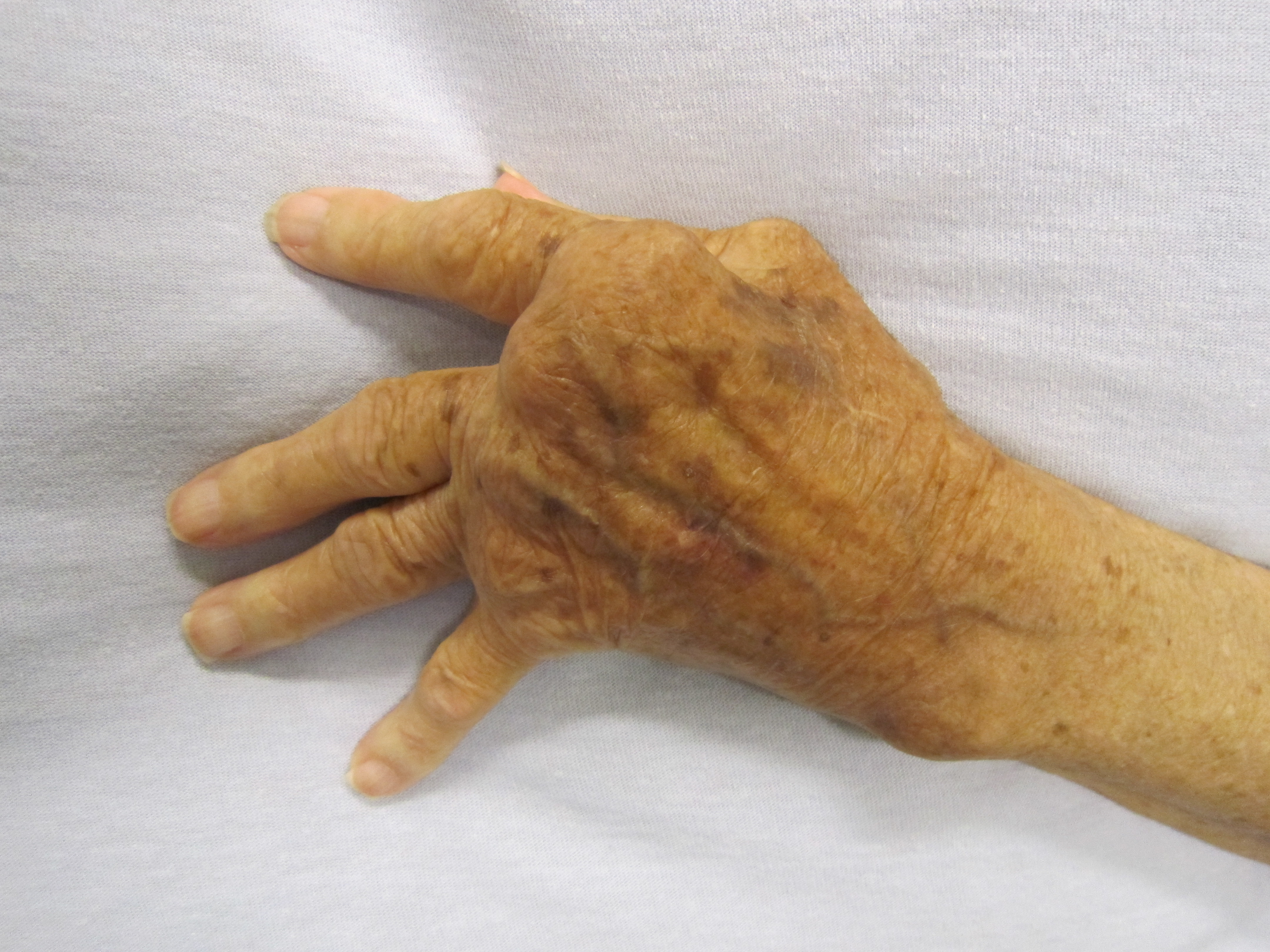
Vignette. A 42-year-old woman has 4 months of symmetric pain and swelling in the MCP and PIP joints of both hands and both wrists, with morning stiffness lasting 2 hours. Exam shows boggy synovitis and early ulnar deviation; the DIP joints are spared. ESR and CRP are elevated.
Most likely diagnosis: Rheumatoid arthritis.
Best initial tests: anti-CCP (most specific) and rheumatoid factor, plus ESR/CRP and X-rays of hands and feet.
Next best step in management: once diagnosed, start methotrexate (first-line DMARD) with folic acid, and bridge with low-dose glucocorticoids for rapid symptom control. Early DMARD therapy prevents irreversible erosions — the goal is treat-to-target (low disease activity or remission).
Diagnosis — antibodies, fluid, imaging
Autoantibodies. Anti-CCP is the most specific (~95%) marker and predicts erosive disease. RF is more sensitive but not specific — also positive in Sjögren, hepatitis C, endocarditis, and SLE. Seronegative RA exists, so diagnosis is clinical using the 2010 ACR/EULAR criteria (joint count, serology, acute-phase reactants, symptom duration ≥6 weeks).
Synovial fluid. Inflammatory: WBC ~2,000–50,000/µL, neutrophil-predominant, cloudy, no crystals, negative Gram stain/culture — obtained mainly to exclude septic and crystal arthritis.
Imaging. Early X-ray shows soft-tissue swelling and juxta-articular osteopenia; later, symmetric joint-space narrowing and marginal erosions (contrast with osteophytes in OA). MRI/ultrasound detect synovitis and erosions earlier and are more sensitive. Obtain cervical flexion–extension films in long-standing RA before intubation.

RA vs osteoarthritis
| Feature | Rheumatoid arthritis | Osteoarthritis |
|---|---|---|
| Mechanism | Autoimmune, inflammatory | Degenerative ("wear and tear") |
| Joints | MCP, PIP, wrists; symmetric; DIP spared | DIP (Heberden), PIP (Bouchard), hips, knees, spine |
| Stiffness | Morning >1 h, better with use | <30 min, worse with use/end of day |
| Labs | ↑ESR/CRP, RF, anti-CCP | Normal inflammatory markers |
| Synovial fluid | Inflammatory (2k–50k WBC, PMNs) | Non-inflammatory (<2000 WBC) |
| X-ray | Osteopenia, marginal erosions, symmetric JSN | Osteophytes, subchondral sclerosis/cysts, asymmetric JSN |
| Nodules | Rheumatoid nodules | Bony Heberden/Bouchard nodes |
Vignette. A 55-year-old woman with RA has persistent moderate disease activity despite 3 months of methotrexate. Her rheumatologist plans to add a TNF-α inhibitor (e.g., etanercept, adalimumab).
Next best step BEFORE starting the biologic: screen for latent tuberculosis (interferon-γ release assay or PPD) and hepatitis B/C; TNF inhibitors can cause reactivation of TB. Update vaccines first and avoid live vaccines during immunosuppression.
High-yield pitfalls:
- Methotrexate is teratogenic — stop it and counsel contraception before conception; it also causes hepatotoxicity and pneumonitis (give folic acid; monitor CBC/LFTs).
- Avoid TNF inhibitors in decompensated heart failure and demyelinating disease.
- Start DMARDs early to prevent erosions; target remission or low disease activity.
- Methotrexate = first-line anchor DMARD; alternatives: leflunomide, sulfasalazine, hydroxychloroquine.
- Bridge with low-dose glucocorticoids for flares; NSAIDs give symptom relief only and do not alter disease course.
- Inadequate response → add a targeted agent, typically a TNF inhibitor first (most long-term data); alternatives include IL-6 inhibitor (tocilizumab), T-cell co-stimulation blocker (abatacept), anti-CD20 (rituximab), or a JAK inhibitor (tofacitinib) — JAK inhibitors carry boxed warnings for thrombosis, MACE, and malignancy.
- Before immunosuppression: screen latent TB and hepatitis B/C; give non-live vaccines.
- Monitor methotrexate: CBC, LFTs, folic acid supplementation; watch for pneumonitis.
- Aggressively manage cardiovascular risk (leading cause of death) and osteoporosis.
Hand deformities
- Boutonnière = "button-hole": PIP flexion + DIP extension (the PIP pokes through like a button).
- Swan-neck: PIP hyperextension + DIP flexion (the neck arches up at the PIP).
Felty syndrome — the triad
- RA + Splenomegaly + Neutropenia ("big spleen, low neutrophils" in seropositive, long-standing RA).
Joint pattern
- RA spares the DIP. DIP involvement instead points to OA (Heberden nodes) or psoriatic arthritis.
Practice MSK now
Board-style questions, spaced-repetition flashcards, and a Socratic AI tutor — free to start.