Pheochromocytoma & Primary Hyperaldosteronism
A boards-focused diagnostic and management comparison of pheochromocytoma (catecholamine excess) and primary hyperaldosteronism/Conn syndrome (autonomous aldosterone), built around the screen → confirm → localize → treat pathway, correct dynamic-test interpretation, and the two classic traps (alpha-before-beta blockade; drawing an ARR without prepping the patient).
Two adrenal causes of secondary hypertension
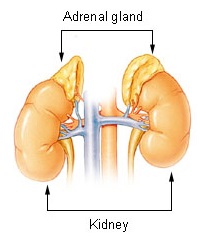
Both diseases are surgically correctable causes of hypertension arising from the adrenal gland — but from different layers with different hormones, so the workup diverges immediately.
- Pheochromocytoma — a catecholamine-secreting tumor of chromaffin cells in the adrenal *medulla* (identical tumors arising in extra-adrenal sympathetic ganglia are paragangliomas). It floods the circulation with epinephrine/norepinephrine.
- Primary hyperaldosteronism (Conn syndrome) — autonomous aldosterone overproduction from the adrenal cortex (zona glomerulosa), independent of the renin–angiotensin system.
The board-tested diagnostic skeleton is the same for both: (1) screen biochemically → (2) confirm with a dynamic/suppression test → (3) localize with imaging (± adrenal vein sampling) → (4) treat — resect a unilateral lesion, medically manage bilateral disease. Getting the order right — biochemistry before imaging — is a recurring trap, because incidental non-functioning adrenal nodules are common and chasing an image first leads you astray.
Highest-yield points
Pheochromocytoma
- Best screening test = plasma free metanephrines (or 24-h urinary fractionated metanephrines). Metanephrines are the COMT-derived O-methylated metabolites produced continuously inside the tumor, so they outperform plasma/urine catecholamines, which are secreted episodically and are easily missed between paroxysms.
- Alpha-blockade must precede beta-blockade — phenoxybenzamine (or doxazosin) for 10–14 days with volume repletion, then add a beta-blocker. A beta-blocker given first causes unopposed alpha vasoconstriction → hypertensive crisis.
- ~40% are hereditary — think MEN2A/2B (RET), VHL, NF1, and SDHx paragangliomas. (The old "10% familial" figure is outdated.)
Primary hyperaldosteronism
- Screen with the aldosterone-to-renin ratio (ARR): high aldosterone with suppressed renin. The suppressed renin is what separates it from secondary hyperaldosteronism (renin high).
- Confirm with a suppression test (oral salt loading or IV saline infusion): aldosterone fails to suppress.
- Lateralize with adrenal vein sampling (AVS) — CT alone is unreliable. Unilateral adenoma → laparoscopic adrenalectomy; bilateral hyperplasia → mineralocorticoid-receptor antagonist (spironolactone or eplerenone).
- Classic triad: resistant hypertension + hypokalemia + metabolic alkalosis (though many patients are now normokalemic at diagnosis).
Recognize it, then pick the next step
Vignette A — Episodic pounding headache, palpitations, and drenching sweats with paroxysmal hypertension; BP spikes on abdominal palpation or at anesthesia induction. → Next step: plasma free metanephrines — not a CT first. If positive, CT/MRI abdomen; if biochemistry is positive but cross-sectional imaging is negative, obtain ¹²³I-MIBG scintigraphy or ⁶⁸Ga-DOTATATE PET.
Vignette B — Treatment-resistant hypertension (uncontrolled on 3 drugs) with spontaneous or diuretic-induced hypokalemia and metabolic alkalosis, no edema. → Next step: aldosterone-to-renin ratio. If the ARR is high, confirm with saline-infusion/oral-salt suppression, then adrenal CT + AVS to lateralize.
Two high-frequency traps:
- Do not start (or lead with) a beta-blocker in suspected pheochromocytoma.
- Do not draw an ARR while the patient is on a mineralocorticoid-receptor antagonist, and correct hypokalemia first — both produce false-negative screening.
| Feature | Pheochromocytoma | Primary hyperaldosteronism (Conn) |
|---|---|---|
| Cell / layer of origin | Chromaffin cells, adrenal medulla | Zona glomerulosa, adrenal cortex |
| Excess hormone | Catecholamines (epi/norepi) → metanephrines | Aldosterone |
| Classic presentation | Episodic headache, palpitations, diaphoresis, paroxysmal HTN | Resistant HTN, hypokalemia, metabolic alkalosis, no edema |
| Electrolytes | Usually normal; may have hyperglycemia | ↓ K⁺, ↑ HCO₃⁻, mildly ↑ Na⁺ |
| Screening test | Plasma free (or 24-h urinary fractionated) metanephrines | Aldosterone-to-renin ratio (↑ aldo, ↓ renin) |
| Confirmatory / dynamic test | Clonidine suppression — metanephrines fail to suppress | Saline infusion / oral salt load — aldosterone fails to suppress |
| Localization | CT/MRI → MIBG or DOTATATE PET if needed | Adrenal CT → adrenal vein sampling |
| Definitive treatment | Alpha-block → beta-block → laparoscopic resection | Unilateral → adrenalectomy; bilateral → spironolactone / eplerenone |
| Key associations | MEN2 (RET), VHL, NF1, SDHx | Bilateral idiopathic hyperplasia (most common); GRA (familial type I) |

Dynamic tests and the mechanisms behind them
Clonidine suppression (pheochromocytoma). Clonidine is a central α₂-agonist that shuts off neurogenic catecholamine release. In healthy people plasma normetanephrine falls; in pheochromocytoma the tumor secretes autonomously, so metanephrines stay elevated — confirming the diagnosis when screening values are borderline.
Suppression tests (primary hyperaldosteronism). A sodium/volume load (oral salt or IV saline) — or a captopril challenge — normally suppresses aldosterone through renin–angiotensin feedback. In primary aldosteronism, secretion is autonomous and fails to suppress.
Why there's little edema — "aldosterone escape." Sustained aldosterone drives early Na⁺/water retention, but the resulting volume expansion triggers pressure natriuresis and atrial natriuretic peptide, resetting to a new steady state with only mild hypernatremia and no edema — while K⁺ and H⁺ wasting continue, producing hypokalemic metabolic alkalosis.
Prep pitfalls for the ARR: correct hypokalemia first (low K⁺ suppresses aldosterone → false negative); keep sodium intake liberal; stop mineralocorticoid-receptor antagonists ~4–6 weeks beforehand. Watch drug effects on the ratio — beta-blockers raise the ARR (false positive), while ACE inhibitors/ARBs and diuretics lower it (false negative). Verapamil, hydralazine, and alpha-blockers are the "clean" agents for BP control during testing.
Memory hooks (only the real ones)
Pheochromocytoma — the "5 P's":
- Pressure (hypertension)
- Pain (headache)
- Palpitations
- Perspiration
- Pallor
"A before B" — give the Alpha-blocker before the Beta-blocker. Reverse the order and you provoke a crisis.
Classic "Rule of 10s" (useful but partly dated): ~10% extra-adrenal, bilateral, malignant, in children, and calcify — but hereditary disease is really ~40%, not 10%.
Primary hyperaldosteronism pattern: aldosterone ↑, renin ↓ (the ratio is the screen). Contrast with secondary hyperaldosteronism (e.g., renal artery stenosis), where both aldosterone and renin are high.
Management pearls to close
Pheochromocytoma — perioperative. After 10–14 days of alpha-blockade with deliberate salt and fluid loading (to refill the chronically vasoconstricted, volume-contracted vascular bed), add a beta-blocker only once alpha-blockade is established, to control reflex tachycardia. Intraoperative hypertensive surges are managed with IV phentolamine or nitroprusside; anticipate post-resection hypotension from the abrupt loss of catecholamines and treat with fluids. Offer germline mutation testing and genetic counseling, and keep lifelong follow-up — histology cannot reliably predict which tumors recur or metastasize.
Primary hyperaldosteronism. A confirmed unilateral source (adenoma or unilateral hyperplasia on AVS) is best treated by laparoscopic adrenalectomy, which reliably corrects hypokalemia and improves — sometimes cures — the hypertension. Bilateral idiopathic hyperplasia is managed medically with a mineralocorticoid-receptor antagonist; eplerenone is preferred when spironolactone's anti-androgen effects (gynecomastia, menstrual irregularity) are limiting. Glucocorticoid-remediable aldosteronism (familial type I) — a chimeric CYP11B1/CYP11B2 gene placing aldosterone synthase under ACTH control — responds to low-dose glucocorticoid.
Practice Endocrine now
Board-style questions, spaced-repetition flashcards, and a Socratic AI tutor — free to start.