Paget Disease, Osteomalacia & Rickets
A Step 1 high-yield lesson contrasting Paget disease (isolated ↑ALP, normal Ca/phosphate, mosaic bone, IV bisphosphonate first-line) with vitamin D–deficient osteomalacia and rickets (↓Ca, ↓phosphate, ↑ALP, ↑PTH via secondary hyperparathyroidism), organized around the Ca/phosphate/ALP/PTH panel with classic radiographic buzzwords and next-best-step decisions. It explicitly notes these metabolic bone diseases lack diagnostic autoantibodies and synovial-fluid findings. Reviewed for clinical accuracy: mechanisms, lab patterns, and first-line management verified; all three Wikimedia Commons image files confirmed to exist.
Three diseases, one lab framework
Paget disease, osteomalacia, and rickets are metabolic bone disorders that Step 1 tests through a calcium / phosphate / ALP / PTH panel plus classic radiographic buzzwords. Note up front: these are diseases of bone remodeling and mineralization — there are no diagnostic autoantibodies and no characteristic synovial-fluid findings (unlike inflammatory arthritis). Diagnosis rests on serum chemistry and imaging.
- Paget disease = focal disordered remodeling: frenzied osteoclastic resorption followed by chaotic osteoblastic overactivity → enlarged but structurally weak "mosaic" (jigsaw) bone. Calcium and phosphate normal; ALP markedly elevated.
- Osteomalacia (adults) and rickets (children) = defective mineralization of osteoid, usually from vitamin D deficiency (or hypophosphatemia). Same pathology; rickets additionally deforms the open growth plate.
The unifying question is always: is calcium normal (Paget) or low with secondary hyperparathyroidism (vitamin D–deficient osteomalacia/rickets)?
- Who/how found: older adults (>55); often asymptomatic, discovered via isolated ↑ALP on routine labs.
- Phases: lytic (osteoclastic) → mixed → sclerotic/blastic → quiescent.
- Symptoms: bone pain, skull enlargement (hats no longer fit), frontal bossing, hearing loss (cochlear/ossicle involvement, ± CN VIII), anterior tibial bowing ("saber shin"), warmth over bone (hypervascular).
- Labs: ↑↑↑ALP and bone-turnover markers; calcium and phosphate normal (Ca can rise with immobilization).
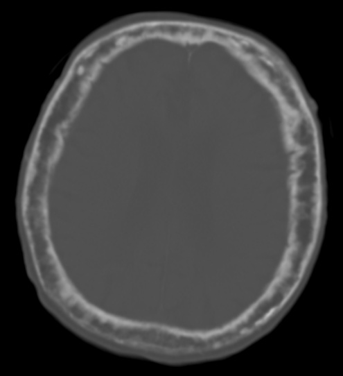
- X-ray buzzwords: "cotton-wool" skull, osteoporosis circumscripta (large lytic skull lesion), "blade-of-grass"/flame-shaped lytic front in long bones, cortical thickening, coarse trabeculae, "picture-frame" vertebra. Bone scan: ↑ uptake.
- Complications: high-output heart failure (AV shunting), osteosarcoma (<1%, feared), pathologic/chalk-stick fractures, secondary osteoarthritis, nerve compression / spinal stenosis.
- Treatment: bisphosphonate (IV zoledronic acid) first-line for symptomatic disease; calcitonin second-line.

- Mechanism: unmineralized osteoid accumulates → soft bone. Most often vitamin D deficiency (poor intake/sunlight, malabsorption, impaired activation in liver/kidney disease); also hypophosphatemia — X-linked hypophosphatemic rickets (FGF23-mediated) and tumor-induced osteomalacia.
- Vitamin D path: skin (7-dehydrocholesterol →UV→ D3) → liver 25-hydroxylase → 25-OH-D (best marker of stores) → kidney 1α-hydroxylase → 1,25-(OH)₂-D (active).
- Labs (vitamin D deficiency): ↓25-OH-D → ↓/low-normal Ca → secondary hyperparathyroidism (↑PTH) → ↓phosphate (phosphaturia) and ↑ALP.
- Rickets (children): bowed legs (genu varum), rachitic rosary (costochondral beading), frontal bossing, craniotabes, widened wrists/ankles, delayed fontanelle closure and dentition; classic in exclusively breastfed infants without vitamin D supplementation.
- Osteomalacia (adults): diffuse bone pain, proximal muscle weakness / waddling gait, fractures; Looser zones (pseudofractures / Milkman lines) on X-ray.
- Treatment: vitamin D + calcium; treat the cause. XLH: phosphate + calcitriol, or burosumab (anti-FGF23).
Vignette: A 72-year-old man reports his hats have grown tight over the past year and notes new hearing loss and dull thigh pain. Labs: ALP markedly elevated; calcium and phosphate normal; PTH normal. Skull X-ray shows patchy "cotton-wool" opacities.
Diagnosis: Paget disease of bone.
Next best step / management:
- Recognize the signature — isolated ↑ALP with normal Ca/phosphate; obtain plain films ± radionuclide bone scan to map disease extent.
- Treat symptomatic disease with a bisphosphonate (IV zoledronic acid).
- Red flag: a sudden increase in pain or a new soft-tissue mass over pagetic bone → suspect osteosarcoma → urgent MRI and biopsy.
Vignette: An 18-month-old who is exclusively breastfed with minimal sun exposure has bowed legs, beading at the costochondral junctions (rachitic rosary), and a delayed-closing anterior fontanelle. Labs: ↓Ca, ↓phosphate, ↑ALP, ↑PTH, ↓25-OH vitamin D. Wrist X-ray shows metaphyseal cupping/fraying with widened growth plates.
Diagnosis: Nutritional (vitamin D–deficiency) rickets.
Next best step:
- Order 25-OH vitamin D (best measure of stores) plus Ca / phosphate / ALP / PTH.
- Treat with vitamin D + calcium repletion; counsel that breastfed infants need 400 IU/day vitamin D.
- If instead labs show normal Ca/PTH with low phosphate and renal phosphate wasting → think X-linked hypophosphatemic rickets, not nutritional deficiency.

Lab pattern comparison (the money table)
| Condition | Ca | Phosphate | ALP | PTH | Classic clue |
|---|---|---|---|---|---|
| Osteoporosis | Normal | Normal | Normal | Normal | Fragility fracture, ↓bone mass, normal labs |
| Paget disease | Normal | Normal | ↑↑↑ | Normal | ↑hat size, hearing loss, mosaic bone |
| Osteomalacia / rickets (vit D def) | ↓ | ↓ | ↑ | ↑ (2°) | Looser zones (adult) / bowed legs (child) |
| Primary hyperparathyroidism | ↑ | ↓ | ↑ | ↑ | Stones, bones, groans; osteitis fibrosa cystica |
| Osteopetrosis | Normal/↓ | Normal | Normal | Normal/↑ | Dense "stone bone," fractures, marrow failure |
- Isolated ↑ALP = Paget (with normal Ca/phosphate) — the single most-tested pearl.
- "Looser zones = loose (soft) bones" → osteomalacia pseudofractures (a.k.a. Milkman lines).
- Vitamin D: "Liver 25, Kidney 1" — liver adds the 25-OH (storage form, best test); kidney adds the 1α-OH to make active 1,25-(OH)₂-D.
- Rachitic rosary + ricket-y wrists → costochondral beading and widened metaphyses in children.
- Paget = "mosaic / jigsaw" histology → disorganized woven + lamellar bone that is big but brittle.
Practice MSK now
Board-style questions, spaced-repetition flashcards, and a Socratic AI tutor — free to start.