Osteoporosis & Metabolic Bone Disease
A high-yield STEP 2 CK lesson on osteoporosis and metabolic bone disease covering DEXA/T-score criteria, screening and FRAX thresholds, a labs-based comparison of osteoporosis vs osteomalacia/Paget/hyperparathyroidism, and a drug table with two exam-style vignettes on fragility fracture and atypical femoral fracture.
What Osteoporosis Is
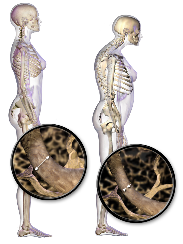
Osteoporosis is reduced bone mass with microarchitectural deterioration and normally mineralized bone, producing skeletal fragility and low-trauma (fragility) fractures — classically vertebral compression, hip (femoral neck), and distal radius (Colles). It is clinically silent until a fracture occurs. The mechanism is imbalanced remodeling: osteoclastic resorption outpaces osteoblastic formation.
Type 1 (postmenopausal) reflects estrogen loss, which unrestrains osteoclasts and preferentially thins trabecular bone (vertebrae, distal radius). Type 2 (senile) is age-related and affects both cortical and trabecular bone.
In primary osteoporosis, serum calcium, phosphate, alkaline phosphatase, and PTH are normal — abnormal values point to a secondary cause (glucocorticoids, hyperthyroidism, primary hyperparathyroidism, hypogonadism, multiple myeloma, malabsorption) or a different metabolic bone disease.
- DEXA of the hip + lumbar spine is the standard for bone mineral density (BMD)
- T-score = SDs vs a young healthy adult; Z-score = SDs vs age-matched peers (use Z-score in premenopausal women, men < 50, and children)
- Normal ≥ -1.0 · Osteopenia -1.0 to -2.5 · Osteoporosis ≤ -2.5; a fragility fracture of the hip or spine is diagnostic regardless of the T-score
- Screen all women ≥ 65 (USPSTF); screen younger postmenopausal women whose fracture risk equals that of a 65-year-old
- FRAX estimates 10-year fracture probability; treat osteopenia when 10-yr hip ≥ 3% or major osteoporotic ≥ 20%
- Risk factors: advancing age, female sex, White/Asian ethnicity, low body weight, early menopause/estrogen deficiency, glucocorticoids (most common secondary cause), smoking, excess alcohol, sedentary lifestyle, low calcium/vitamin D, family history

Metabolic Bone Disease at a Glance
| Disease | Ca | Phosphate | ALP | PTH | Clues |
|---|---|---|---|---|---|
| Osteoporosis | Normal | Normal | Normal | Normal | ↓ mass, normal mineralization |
| Osteomalacia / rickets (vit D def.) | Low–normal | Low | High | High | Looser pseudofractures, bowing |
| Paget disease of bone | Normal | Normal | Very high (isolated) | Normal | Bone pain, ↑ hat size, CN VIII hearing loss, high-output HF |
| Primary hyperparathyroidism | High | Low | High | High | Stones, bones, groans; osteitis fibrosa cystica |
| Osteopetrosis | Normal–low | Normal | Normal | Normal | Dense, brittle bone; marrow failure |
Vignette: A 68-year-old woman develops acute mid-back pain after lifting a laundry basket. She underwent menopause at 46, smokes, and is thin. Spine X-ray shows a T8 vertebral compression fracture with no significant trauma.
Diagnosis: Osteoporotic fragility fracture — a low-trauma vertebral (or hip) fracture is itself diagnostic of osteoporosis, independent of BMD.
Next steps:
- Obtain DEXA for baseline BMD and to monitor therapy
- Lab workup for secondary causes: CBC, calcium/phosphate, 25-OH vitamin D, PTH, TSH, creatinine, and SPEP/UPEP if myeloma is suspected
- Start calcium + vitamin D and a first-line oral bisphosphonate (alendronate)
- Provide analgesia and encourage early mobilization; avoid prolonged bed rest
Treatment Principles
Who to treat: T-score ≤ -2.5, any hip/vertebral fragility fracture, or osteopenia with high FRAX risk.
All patients: weight-bearing exercise, smoking/alcohol reduction, fall-proofing, calcium ~1200 mg/day and vitamin D 800–1000 IU/day. Correct vitamin D deficiency before starting an antiresorptive to avoid precipitating hypocalcemia.
First-line = bisphosphonates (antiresorptive). Oral agents must be taken on an empty stomach with a full glass of water while remaining upright ≥ 30 minutes to prevent pill esophagitis; avoid if CrCl < 30–35 or uncorrected hypocalcemia. After 3–5 years, lower-risk patients may take a drug holiday.
Anabolic agents (teriparatide, romosozumab) are preferred for very severe or very-high-risk disease; they build new bone and are then followed by an antiresorptive to preserve the gains.
Drugs for Osteoporosis
| Drug (class) | Mechanism | Key adverse effects / notes |
|---|---|---|
| Alendronate, risedronate, IV zoledronate (bisphosphonate) | Inhibit osteoclast resorption (↓ farnesyl-PP synthase) | First-line; pill esophagitis, atypical (subtrochanteric) femur fracture, osteonecrosis of jaw; IV → acute flu-like reaction |
| Denosumab (RANKL mAb) | Blocks RANKL → ↓ osteoclast formation | Usable in CKD; do not stop abruptly (rebound vertebral fractures); hypocalcemia, ONJ |
| Raloxifene (SERM) | Estrogen agonist on bone | ↓ vertebral fracture + breast-cancer risk; ↑ VTE, hot flashes; no hip-fracture benefit |
| Teriparatide / abaloparatide (PTH / PTHrP analog) | Intermittent PTH-receptor agonism → osteoblast (anabolic) | Severe disease, ≤ 2 yr; avoid in Paget disease or prior bone radiation |
| Romosozumab (anti-sclerostin mAb) | ↑ formation + ↓ resorption | Anabolic; cardiovascular (MI/stroke) boxed warning |
Vignette: A 74-year-old woman on alendronate for 7 years reports several weeks of dull right thigh/groin pain. After a minor stumble she sustains a fracture; X-ray shows a transverse fracture of the subtrochanteric femur with focal lateral cortical thickening (beaking).
Diagnosis: Atypical femoral fracture — a recognized complication of long-term bisphosphonate (or denosumab) therapy. Prodromal thigh/groin pain and a transverse subtrochanteric/diaphyseal pattern are the tip-offs; it is often bilateral.
Next steps:
- Stop the bisphosphonate
- Image the contralateral femur (frequently involved)
- Orthopedic fixation; ensure adequate calcium/vitamin D
- Contrast with osteonecrosis of the jaw, the other classic bisphosphonate complication (often after dental extraction)
Practice Endocrine now
Board-style questions, spaced-repetition flashcards, and a Socratic AI tutor — free to start.