Osteoarthritis, Gout & Pseudogout
A board-focused MSK/rheumatology lesson contrasting osteoarthritis (degenerative), gout (MSU crystals), and pseudogout (CPPD crystals) across pathophysiology, presentation, synovial fluid and imaging, and next-best-step management. Emphasizes arthrocentesis to exclude septic arthritis, birefringence patterns, negative autoantibodies, and the rule against starting urate-lowering therapy during an acute flare.
Three Arthritides, One Aspirating Needle
Osteoarthritis (OA), gout, and pseudogout are the classic non-autoimmune joint diseases—boards contrast them with RA and SLE, because here RF, anti-CCP, and ANA are negative.
OA is degenerative ("wear-and-tear"): chondrocyte-driven breakdown of articular cartilage outpaces repair, exposing subchondral bone. It is mechanical, not primarily inflammatory (synovial WBC <2,000).
Gout and pseudogout are crystal-induced. In gout, hyperuricemia drives monosodium urate (MSU) deposition; in pseudogout, calcium pyrophosphate dihydrate (CPPD) deposits. Both activate the NLRP3 inflammasome → IL-1β, producing an acute inflammatory monoarthritis (WBC 2,000–50,000).
The unifying board skill: an acutely hot, swollen joint demands arthrocentesis—you must exclude septic arthritis and identify crystals before committing to therapy. A normal serum urate never excludes gout during a flare.
- Who/where: older, obese, prior joint injury; weight-bearing joints (knee, hip), DIP (Heberden nodes), PIP (Bouchard nodes), first CMC (thumb base), spine
- Pain pattern: worse with use, better with rest; morning stiffness <30 min; no systemic symptoms
- Spares the MCP and wrist—those point to RA (or hemochromatosis, which favors the 2nd–3rd MCPs)
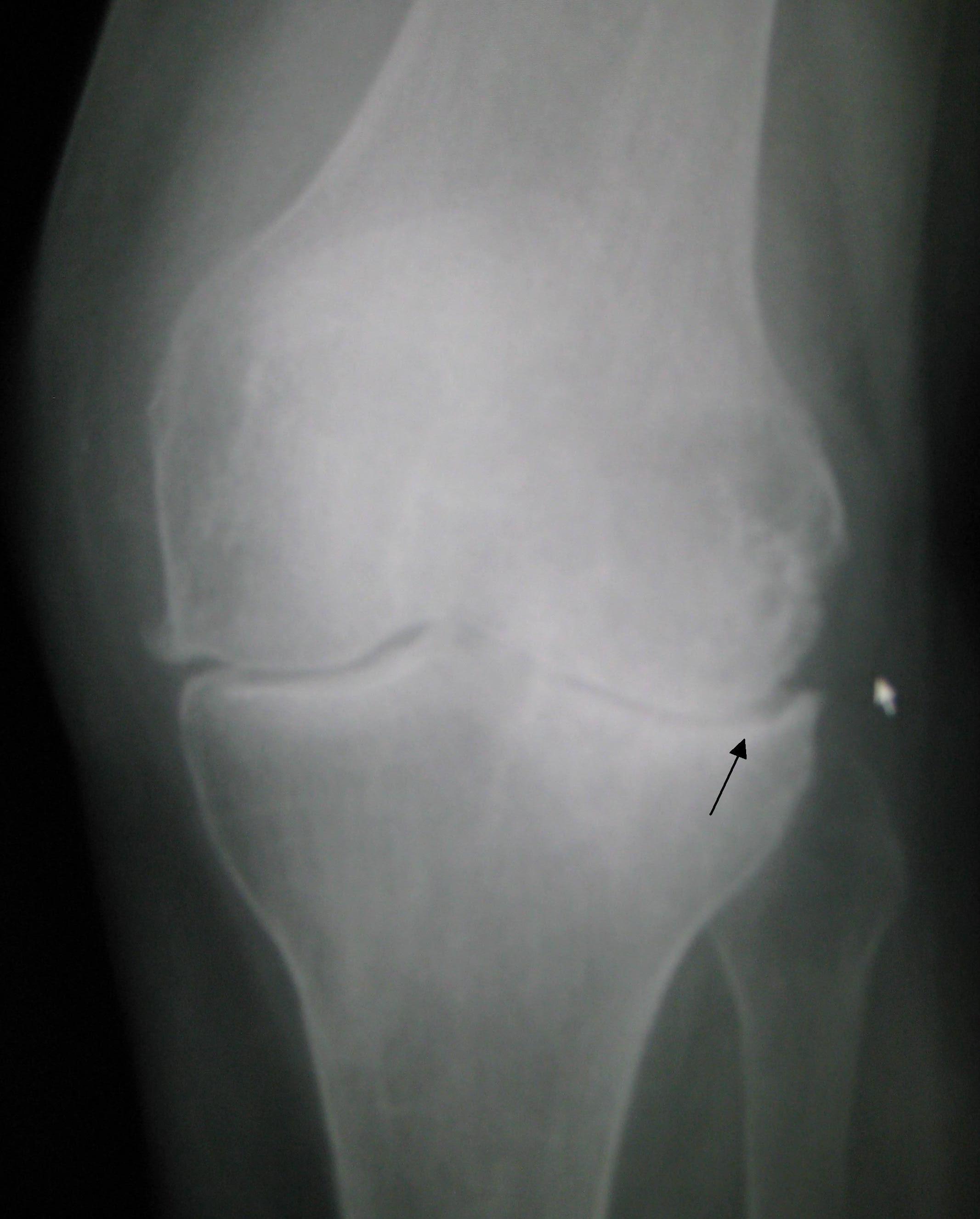
- X-ray "LOSS": Loss of joint space (asymmetric), Osteophytes, Subchondral sclerosis, Subchondral cysts
- Synovial fluid: non-inflammatory, clear, WBC <2,000
- Management ladder: 1) exercise, weight loss, PT (first-line); 2) topical NSAIDs (hand/knee) → oral NSAIDs; 3) intra-articular steroids; duloxetine as adjunct; 4) joint replacement for refractory end-stage disease
- Not recommended: opioids, glucosamine/chondroitin (no proven benefit)
A 64-year-old obese woman reports bilateral knee and hand pain that worsens through the day and eases with rest. Exam: bony enlargement of the DIP joints, <15 min of morning stiffness, no warmth or synovitis. RF and anti-CCP are negative.
- Diagnosis: osteoarthritis; DIP nodules = Heberden nodes.
- Best initial test: none required—OA is clinical; a weight-bearing X-ray (LOSS findings) confirms if uncertain.
- Next best step (management): structured exercise + weight loss, then topical/oral NSAIDs.
- Trap: DIP involvement with negative serologies argues against RA, which favors MCP/PIP/wrist with +anti-CCP and >30–60 min morning stiffness.
- Cause: hyperuricemia from underexcretion (~90%) (CKD, thiazide/loop diuretics, alcohol) or overproduction (~10%) (tumor lysis, myeloproliferative disease, Lesch-Nyhan = HGPRT deficiency)
- Triggers: alcohol (esp. beer), red meat, seafood, fructose, dehydration, surgery
- Classic: acute podagra (first MTP)—red, hot, exquisitely tender, often starting at night; chronic → tophi
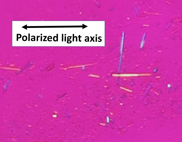
- Crystals: needle-shaped, negatively birefringent (yellow when parallel to the compensator)
- X-ray: punched-out / "rat-bite" erosions with overhanging edges; joint space preserved until late
- Acute Rx: NSAIDs, colchicine, or corticosteroids (systemic or intra-articular)
- Do NOT start or stop urate-lowering therapy (ULT) during a flare—but continue it if the patient is already on it
- Chronic ULT (target urate <6 mg/dL): allopurinol (xanthine oxidase inhibitor) first-line → febuxostat; probenecid if underexcretor with good renal function and no stones. Indicated for ≥2 flares/yr, tophi, or urate stones
- Give colchicine/NSAID prophylaxis when starting ULT; allopurinol + azathioprine/6-MP = toxic (blocks their metabolism → myelosuppression)
A 55-year-old man wakes with an acutely swollen, red, exquisitely tender first MTP joint after a steak-and-beer dinner. Temp 37.8°C. Serum urate is normal.
- Next best step (diagnosis): arthrocentesis with synovial fluid analysis—you must exclude septic arthritis.
- Findings: WBC ~20,000; needle-shaped, negatively birefringent crystals → gout.
- Next best step (treatment): NSAIDs, colchicine, or corticosteroids. If CKD or on anticoagulation, favor intra-articular or oral steroids over NSAIDs.
- Do NOT start allopurinol now—initiating ULT mid-flare can worsen it; begin weeks later under prophylaxis.
- Key point: a normal serum urate does not exclude gout during an acute attack.
- Deposits: calcium pyrophosphate dihydrate (CPPD)
- Who/where: older adults; knee (most common) and wrist; acute mono/oligoarthritis that mimics gout
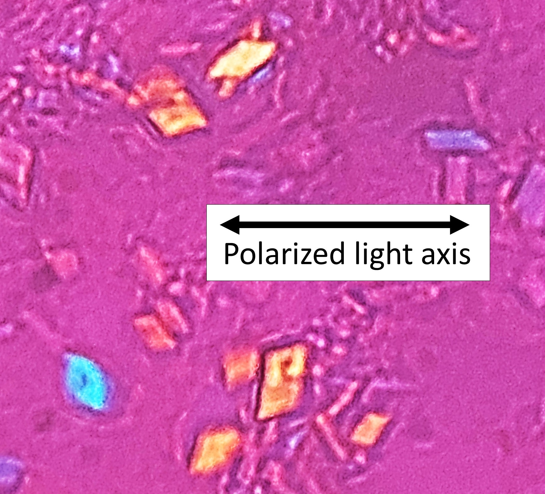
- Crystals: rhomboid-shaped, positively birefringent (blue when parallel to the compensator)
- Imaging: chondrocalcinosis—linear calcification of cartilage (knee menisci, wrist triangular fibrocartilage)
- Secondary causes—the "4 H's": Hemochromatosis, Hyperparathyroidism, Hypophosphatasia, Hypomagnesemia. Work these up in the young or recurrent case
- Management: same as acute gout—NSAIDs, colchicine, or intra-articular/systemic steroids; treat the underlying disorder. No urate-lowering therapy; colchicine prophylaxis for frequent attacks
A 72-year-old presents with an acutely swollen, warm knee. Knee X-ray shows linear calcification within the meniscal cartilage.
- Diagnosis clue: chondrocalcinosis → suspect CPPD (pseudogout).
- Next best step: arthrocentesis—fluid shows rhomboid, positively birefringent crystals; still exclude infection.
- Treatment: intra-articular steroids or NSAIDs/colchicine.
- If the patient is young or has recurrent disease: screen for the 4 H's—iron studies (hemochromatosis), Ca/PTH (hyperparathyroidism), Mg.
- Buzzword link: new CPPD + skin bronzing + diabetes → hemochromatosis.
- OA on X-ray = "LOSS": Loss of joint space, Osteophytes, Subchondral sclerosis, Subchondral cysts
- Gout crystals — the two N's: Needle-shaped and Negatively birefringent
- Birefringence colors: gout (MSU, negative) = yellow when parallel to the compensator, blue when perpendicular; pseudogout (CPPD, positive) = blue when parallel, yellow when perpendicular
- CPPD secondary causes = "4 H's": Hemochromatosis, Hyperparathyroidism, Hypophosphatasia, Hypomagnesemia
Comparison: OA vs Gout vs Pseudogout
| Feature | Osteoarthritis | Gout | Pseudogout (CPPD) |
|---|---|---|---|
| Mechanism | Cartilage degeneration | MSU crystals (hyperuricemia) | CPPD crystals |
| Classic joints | Knee, hip, DIP/PIP, 1st CMC | 1st MTP (podagra), midfoot | Knee, wrist |
| Crystal shape | None | Needle | Rhomboid |
| Birefringence | — | Negative (yellow ∥) | Positive (blue ∥) |
| Synovial WBC | <2,000 (non-inflammatory) | 2,000–50,000 | 2,000–50,000 |
| Imaging | LOSS; osteophytes | Punched-out erosions, overhanging edges | Chondrocalcinosis |
| Autoantibodies | Negative | Negative | Negative |
| Acute Rx | PT, weight loss, NSAIDs | NSAIDs / colchicine / steroids | NSAIDs / colchicine / steroids |
Practice MSK now
Board-style questions, spaced-repetition flashcards, and a Socratic AI tutor — free to start.