Mesenteric Ischemia & Appendicitis
A Step 2 CK high-yield lesson pairing mesenteric ischemia (its four subtypes plus ischemic colitis) with acute appendicitis, driving each pathophysiology → presentation → diagnosis → NEXT-BEST-STEP with classic vignette buzzwords, imaging/lab findings, and comparison tables.
Two Faces of the Acute Surgical Abdomen
Both mesenteric ischemia and acute appendicitis begin as visceral processes that turn into surgical emergencies once the bowel wall is threatened. Mesenteric ischemia is a vascular catastrophe — the tested clue is pain out of proportion to a benign abdominal exam in an older patient with atrial fibrillation or atherosclerosis. Appendicitis is an obstructive one — luminal blockage (an appendicolith/fecalith in adults, lymphoid hyperplasia in children) drives distension, ischemia, and eventual perforation.
On Step 2 CK both reward the same skill: matching the vignette's onset, risk factors, and imaging to the single best next step. Delay kills bowel, so the tested instinct is to resuscitate, image with CT angiography (ischemia) or CT with contrast / ultrasound (appendicitis), and involve surgery before transmural infarction or perforation and peritonitis develop.
- Acute mesenteric ischemia (AMI) = small-bowel emergency; hallmark is pain out of proportion to exam.
- SMA embolism (~50%, most common) — cardiac source (atrial fibrillation, recent MI with LV mural thrombus); sudden, severe pain.
- SMA thrombosis (~15–25%) — on pre-existing atherosclerosis; often preceded by chronic postprandial pain, food fear, and weight loss.
- NOMI (non-occlusive) — low-flow/splanchnic vasoconstriction: shock, vasopressors, digoxin, cocaine.
- Mesenteric venous thrombosis — hypercoagulable states / portal HTN; younger patient, more subacute course; treat with anticoagulation.
- Labs: leukocytosis; elevated lactate + anion-gap metabolic acidosis signals infarction — a late sign. Normal labs do NOT exclude early AMI.
- Best test: CT angiography of the abdomen (catheter angiography is the historical gold standard).
- Peritoneal signs → straight to exploratory laparotomy. Otherwise: IV fluids, broad-spectrum antibiotics, heparin, and revascularization/embolectomy (surgical or endovascular).
Vignette: A 74-year-old man with atrial fibrillation (not anticoagulated) develops sudden, severe, diffuse abdominal pain. Exam: a soft abdomen with only mild tenderness despite obvious distress. Lactate 4.8, WBC 18k, anion-gap metabolic acidosis.
- Diagnosis: Acute mesenteric ischemia from SMA embolism (AFib → cardioembolism; pain out of proportion to exam).
- Best confirmatory test: CT angiography of the abdomen/pelvis.
- Next steps: IV fluids, broad-spectrum antibiotics, systemic anticoagulation (heparin), and urgent surgery/IR consult for embolectomy or revascularization. Avoid vasoconstrictors (they worsen splanchnic flow).
- If rebound/guarding (peritonitis): go directly to exploratory laparotomy to resect infarcted bowel — do not delay for more imaging.
Intestinal Ischemia — 3-Way Comparison
| Feature | Acute Mesenteric Ischemia | Ischemic (Colonic) Colitis | Chronic Mesenteric Ischemia |
|---|---|---|---|
| Vessel / bowel | SMA → small bowel | Watershed colon: splenic flexure (Griffith), rectosigmoid (Sudeck) | ≥2 of celiac / SMA / IMA |
| Classic patient | AFib, atherosclerosis | Elderly, hypotension, post-AAA repair | Diffuse atherosclerosis, smoker |
| Pain | Severe, out of proportion | Crampy LLQ | Postprandial ("intestinal angina") + food fear, weight loss |
| Bleeding | Minimal early | Hematochezia / bloody diarrhea | None |
| Best test | CT angiography | Colonoscopy (CT: thumbprinting, wall thickening) | CTA / MRA |
| Management | Anticoag + revascularization; laparotomy if infarct | Supportive (bowel rest, fluids, abx); usually self-limited | Angioplasty/stent or surgical bypass |
- Pathophysiology: luminal obstruction → distension → venous congestion → ischemia → perforation. Appendicolith/fecalith in adults; lymphoid hyperplasia in children/young adults; peak in teens–20s.
- Classic course: vague periumbilical pain (visceral, T10) that migrates to the RLQ / McBurney point (parietal peritoneum), plus anorexia, nausea, low-grade fever.
- Signs: Rovsing (LLQ press → RLQ pain), psoas (retrocecal appendix), obturator (pelvic appendix), rebound/Blumberg.
- Labs: mild leukocytosis with left shift.
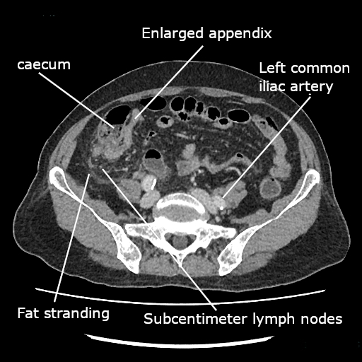
- Imaging: CT abdomen/pelvis with contrast = most accurate in adults (appendix >6 mm, wall thickening, fat stranding, appendicolith). Ultrasound first-line in children & pregnancy (no radiation); MRI if US equivocal in pregnancy.
- Treatment: appendectomy (laparoscopic) + preoperative antibiotics.

Vignette: A 24-year-old woman at 20 weeks' gestation has 1 day of periumbilical pain now localized to the right side, with anorexia and nausea. Temp 38.0°C, right lower/flank tenderness.
- Best initial imaging: graded-compression ultrasound (avoids fetal radiation). MRI without contrast if US is nondiagnostic. Avoid CT.
- Diagnosis: acute appendicitis — the most common non-obstetric surgical emergency in pregnancy; the gravid uterus can displace the appendix cephalad.
- Management: prompt laparoscopic appendectomy + preoperative antibiotics; delay raises perforation and fetal-loss risk.
- Perforation with a walled-off abscess (any patient): CT-guided percutaneous drainage + antibiotics, then interval appendectomy ~6–8 weeks later — not immediate surgery.
MANTRELS captures the classic Alvarado score for appendicitis:
- M — Migration of pain to the RLQ
- A — Anorexia
- N — Nausea / vomiting
- T — Tenderness in the RLQ (2 points)
- R — Rebound tenderness
- E — Elevated temperature
- L — Leukocytosis (2 points)
- S — Shift of WBCs to the left (left shift)
RLQ tenderness and leukocytosis are worth 2 points each (total = 10). A score ≥7 suggests appendicitis (favor appendectomy / surgical consult); 5–6 warrants observation or imaging; ≤4 makes it unlikely.
- "Pain out of proportion" + AFib → acute mesenteric ischemia until proven otherwise → get CT angiography; don't wait on lactate.
- A normal lactate does NOT rule out early mesenteric ischemia.
- Bloody diarrhea + crampy LLQ pain in an elderly or post-AAA-repair patient → ischemic colitis (→ colonoscopy), not AMI.
- Postprandial pain + weight loss + food fear → chronic mesenteric ischemia → elective CTA + revascularization.
- Pregnant patient or child with RLQ pain → ultrasound first, never start with CT.
- Textbook appendicitis in a healthy young man may go straight to appendectomy; reserve CT for equivocal cases, and image women to exclude gynecologic causes (ectopic, ovarian torsion).
- Appendiceal phlegmon / abscess → antibiotics ± drainage + interval appendectomy, not immediate operation.
Practice GI now
Board-style questions, spaced-repetition flashcards, and a Socratic AI tutor — free to start.