Malabsorption Syndromes
A boards-focused walkthrough of malabsorption: three-phase pathophysiology, deficiency-by-location clues, D-xylose logic, and the classic causes (celiac, Whipple, tropical sprue, pancreatic insufficiency, SIBO, abetalipoproteinemia) with next-best-step diagnosis and cause-directed management.
Overview & Pathophysiology
Malabsorption is failure to absorb nutrients across the small-bowel mucosa, distinguished from maldigestion (failure to break nutrients down) — boards often blend the two. Reason in three phases:
- Luminal (digestion): pancreatic enzymes + bile acids. Fails in chronic pancreatitis, cystic fibrosis, cholestasis, and bacterial overgrowth (deconjugates bile salts).
- Mucosal (absorptive): enterocyte brush border + villi. Fails in celiac, tropical sprue, Whipple, lactase deficiency, Crohn.
- Post-mucosal (transport): the enterocyte can't export absorbed lipid, or lymphatics are obstructed. Fails in abetalipoproteinemia (MTP defect → no chylomicron assembly/export) and intestinal lymphangiectasia (blocked lacteals).
The hallmark of fat malabsorption is steatorrhea: bulky, greasy, foul-smelling, pale stools that float and are hard to flush. Because dietary fat carries the fat-soluble vitamins, chronic malabsorption produces ADEK deficiency plus weight loss despite adequate intake. Your board task: use the site of injury and the specific deficiency to name the disease and choose the next best diagnostic step.
- Steatorrhea: bulky, greasy, foul, floating, pale, hard-to-flush stools; weight loss despite normal/increased intake
- Osmotic diarrhea (carbohydrate malabsorption): improves with fasting, ↑ stool osmotic gap, low stool pH
- Deficiency localizes the lesion:
- Duodenum / proximal jejunum → iron (microcytic anemia) + folate → celiac
- Terminal ileum → B12 + bile salts → Crohn, ileal resection
- SIBO is the exception to "location": bacteria synthesize folate (↑) but consume B12 (↓) — low B12 with a normal ileum
- Fat-soluble vitamin signs (ADEK): A night blindness; D hypocalcemia/osteomalacia, tetany; E ataxia, peripheral neuropathy, hemolytic anemia; K ↑ PT/INR, easy bruising
- Protein loss → hypoalbuminemia → edema; Ca/Mg loss → tetany
- Children: failure to thrive, abdominal distension, muscle wasting after weaning onto cereals (celiac)
Comparing the Classic Causes
| Disease | Mechanism / buzzword | Distinctive finding | Treatment |
|---|---|---|---|
| Celiac | Gliadin autoimmunity, HLA-DQ2/DQ8 | Anti-tTG IgA; villous atrophy + crypt hyperplasia + IELs; dermatitis herpetiformis | Lifelong gluten-free diet |
| Tropical sprue | Post-tropical travel, ?infectious | Affects whole small bowel → folate (early) + B12 (late), megaloblastic anemia | Tetracycline + folate (3–6 mo) |
| Whipple | Tropheryma whipplei | PAS+ foamy macrophages; older man, arthralgias, CNS/cardiac | Ceftriaxone → TMP-SMX ~1 yr |
| Chronic pancreatitis | ↓ enzymes (alcohol; CF in kids) | Normal D-xylose, low fecal elastase, pancreatic calcifications | Enzyme replacement |
| Lactase deficiency | Brush-border enzyme loss | Symptoms after dairy, normal biopsy, ↑ osmotic gap, +H₂ breath test | Avoid lactose ± lactase |
| SIBO | Overgrowth from stasis/anatomy | ↑ folate, ↓ B12; abnormal D-xylose that corrects with antibiotics | Rifaximin; fix stasis |
| Abetalipoproteinemia | MTP loss → no chylomicrons | Infant FTT, acanthocytes, near-absent LDL, vitamin E neuro | Fat-soluble vitamins (esp. E), restrict LCFA / use MCT |
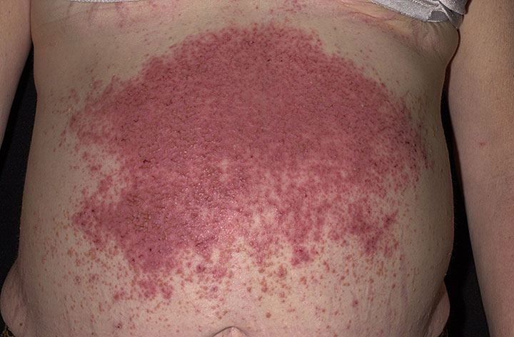
Vignette: A 32-year-old woman has 6 months of bloating and loose, pale stools. Labs show microcytic anemia unresponsive to oral iron. She has an intensely pruritic, grouped vesicular rash on the extensor elbows and knees.
Diagnosis: Celiac disease. The rash is dermatitis herpetiformis (granular IgA deposits at the dermal papillae tips).
Next best step:
- Serology first, while still eating gluten: IgA anti-tissue transglutaminase (tTG) plus total IgA — selective IgA deficiency is associated and causes false negatives, so order IgG-based tTG/DGP if IgA is low.
- Confirm with duodenal biopsy: villous atrophy, crypt hyperplasia, intraepithelial lymphocytosis. Damage is most severe proximally (highest gluten load); take ≥4 biopsies from the distal duodenum plus the bulb because involvement can be patchy.
- Treat: lifelong gluten-free diet; dapsone for the rash.
Board trap: starting a gluten-free diet before testing normalizes serology and histology → test first, then treat.
- Confirm fat malabsorption: 72-hr fecal fat >7 g/day (gold standard); qualitative Sudan III stain of stool
- D-xylose absorption test localizes the defect (xylose is absorbed passively, needs no pancreatic enzymes):
- Low urinary/serum xylose → mucosal disease (celiac, tropical sprue) or SIBO (gut bacteria ferment it → normalizes after antibiotics)
- Normal xylose with high fecal fat → pancreatic exocrine insufficiency (mucosa intact)
- Fecal elastase-1 low → exocrine pancreatic insufficiency
- Celiac: IgA anti-tTG + total IgA → duodenal biopsy to confirm
- Hydrogen breath test: positive in lactase deficiency (lactose substrate) and SIBO (glucose/lactulose)
- Biopsy buzzwords: PAS+ foamy macrophages = Whipple; lipid-laden enterocytes = abetalipoproteinemia; dilated lacteals = lymphangiectasia

Fat-soluble vitamins lost in steatorrhea — "ADEK": A, D, E, K ride with dietary fat, so they drop first in fat malabsorption.
Whipple → "foamy like whipped cream": the lamina propria is packed with PAS-positive foamy macrophages; organism = *Tropheryma whipplei*.
Celiac biopsy triad — "flat, deep, angry": flat mucosa (villous atrophy), deep crypts (crypt hyperplasia), angry lymphocytes (intraepithelial lymphocytosis).
Vignette: A 55-year-old man has several years of migratory arthralgias, then develops weight loss, chronic diarrhea, low-grade fever, and lymphadenopathy. Over months he becomes forgetful with abnormal eye movements and a new heart murmur. Duodenal biopsy shows PAS-positive foamy macrophages in the lamina propria.
Diagnosis: Whipple disease (Tropheryma whipplei) — classically a middle-aged white man; arthralgias precede GI symptoms by years, with systemic cardiac (culture-negative endocarditis), neurologic, and articular involvement. Oculomasticatory myorhythmia (rhythmic eye + jaw movements) is pathognomonic.
Next best step / treatment:
- Confirm with PAS stain ± PCR for T. whipplei.
- Induction: IV ceftriaxone (or penicillin G) ~2 weeks.
- Maintenance: oral TMP-SMX for ~1 year — crosses the blood-brain barrier to clear the CNS reservoir; relapse is common and often neurologic.

- Cause-directed therapy:
- Celiac → lifelong gluten-free diet; replete iron, folate, B12, vitamin D, calcium; DEXA for bone loss
- Pancreatic insufficiency → pancreatic enzyme replacement + fat-soluble vitamins
- SIBO → rifaximin, correct underlying stasis, replete B12
- Tropical sprue → tetracycline + folate; Whipple → ceftriaxone then TMP-SMX
- Lactase deficiency → avoid lactose ± lactase; abetalipoproteinemia / lymphangiectasia → MCT oil (absorbed directly into portal blood, bypassing lymphatics) + fat-soluble vitamins (esp. E)
- Red flags / complications:
- Celiac not responding to a strict gluten-free diet → refractory celiac, enteropathy-associated T-cell lymphoma (EATL), small-bowel adenocarcinoma
- Untreated → osteoporosis, vitamin K coagulopathy, hypocalcemic tetany
Practice GI now
Board-style questions, spaced-repetition flashcards, and a Socratic AI tutor — free to start.