Inflammation & Wound Healing
Inflammation is the body's protective response to injury — acute (neutrophil-driven, vascular) versus chronic (macrophage-driven, fibrotic) — followed by wound healing through defined phases. This lesson locks down the leukocyte adhesion cascade, chemical mediators and their drug targets, granulomatous/adhesion defects, collagen chemistry, and the classic healing failures boards love to test.
The big picture: injury → inflammation → repair
Inflammation is the body's stereotyped, protective response to injury or infection, meant to dilute, destroy, and wall off the offending agent and set the stage for repair. Acute inflammation is rapid in onset (minutes to hours), dominated by neutrophils, and driven by vascular changes plus preformed or rapidly synthesized mediators; it either resolves, forms an abscess, or progresses. Chronic inflammation is persistent (weeks to months), dominated by macrophages, lymphocytes, and plasma cells, and marked by simultaneous tissue destruction, angiogenesis, and fibrosis. Once the noxious stimulus is cleared, wound healing restores tissue by regeneration and/or scarring — a program boards test through its phases, growth factors, collagen chemistry, and characteristic failures.
- Cardinal signs: rubor (redness), calor (heat), tumor (swelling), dolor (pain), functio laesa (loss of function).
- Vascular sequence: transient vasoconstriction → vasodilation (histamine, NO, prostaglandins) → increased vascular permeability → stasis → margination.
- Leukocyte cascade: margination → rolling (selectins: E-/P-selectin binding sialyl-Lewis X) → tight adhesion (integrins: LFA-1 / Mac-1 → ICAM-1 ) → transmigration/diapedesis (PECAM-1 / CD31) → chemotaxis.
- Neutrophil chemotactic factors: C5a, IL-8, LTB4, bacterial N-formylmethionine (fMLP), kallikrein.
- Opsonins: IgG and C3b enhance phagocytosis.
- Respiratory burst: NADPH oxidase → superoxide → (SOD) → H2O2 → (myeloperoxidase) → HOCl (bleach) — the O2-dependent killing system.
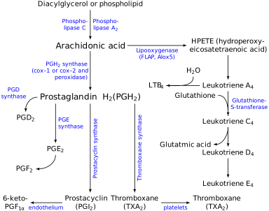
- Key mediators: histamine (vasodilation, permeability), PGE2 (pain, fever, vasodilation), LTB4 (chemotaxis), LTC4/D4/E4 (bronchospasm, permeability, vasoconstriction), bradykinin (pain), C3a/C5a (anaphylatoxins).
- Arachidonic-acid drug targets (high-yield): corticosteroids inhibit phospholipase A2 (↓ all eicosanoids); NSAIDs/aspirin inhibit COX (aspirin = irreversible); zileuton inhibits 5-lipoxygenase; montelukast/zafirlukast block the leukotriene (CysLT1) receptor.
- Acute-phase reactants (IL-6-driven, hepatic): ↑ CRP, fibrinogen, ferritin, hepcidin, serum amyloid A; ↓ albumin, transferrin.
Acute vs chronic inflammation
| Feature | Acute | Chronic |
|---|---|---|
| Onset / duration | Minutes–hours; lasts days | Days–weeks–months |
| Dominant cells | Neutrophils early; macrophages later | Macrophages, lymphocytes, plasma cells |
| Hallmarks | Vascular changes, exudate, edema | Destruction + angiogenesis + fibrosis |
| Key mediators | Histamine, complement, eicosanoids, kinins | IFN-γ, TNF-α, IL-1; growth factors |
| Special pattern | Serous / fibrinous / purulent | Granulomatous (epithelioid + giant cells) |
| Outcome | Resolution, abscess, or → chronic | Scar, granuloma, AA amyloidosis |
PRISH captures the classic signs of acute inflammation:
- Pain (dolor) — bradykinin, PGE2
- Redness (rubor) — vasodilation
- Immobility / loss of function (functio laesa)
- Swelling (tumor) — increased vascular permeability
- Heat (calor) — vasodilation
- Leukocyte adhesion deficiency type 1 (LAD-1): defective β2-integrin (CD18) → leukocytes cannot firmly adhere or transmigrate. Vignette: delayed umbilical cord separation (>30 days), recurrent skin/mucosal infections without pus, and striking neutrophilia (neutrophils stuck in blood).
- Chédiak-Higashi syndrome: LYST mutation → defective phagolysosome fusion and microtubule function. Vignette: partial albinism, recurrent pyogenic infections, peripheral neuropathy, and giant granules in neutrophils on smear.
- Chronic granulomatous disease (CGD): NADPH oxidase defect (often X-linked). Recurrent infections with catalase-positive organisms (S. aureus, Serratia, B. cepacia, Aspergillus, Nocardia). Next step / diagnosis: abnormal dihydrorhodamine (DHR) flow cytometry and a negative nitroblue tetrazolium (NBT) test.
- Granuloma pearl: Th1 → IFN-γ activates macrophages into epithelioid cells; TNF-α maintains the granuloma — so anti-TNF therapy can reactivate latent TB.
Wound healing — regenerate or scar?
Whether tissue regenerates or scars depends on the proliferative capacity of its cells and the integrity of the underlying ECM scaffold. Labile tissues (epidermis, GI and respiratory epithelium, bone marrow, germ cells) cycle continuously and regenerate readily. Stable tissues (hepatocytes, proximal renal tubules) sit quiescent in G0 but re-enter the cycle after injury — the basis of liver regeneration. Permanent tissues (neurons, cardiac and skeletal muscle) cannot effectively divide and heal by scar (fibrosis). Cutaneous healing runs in three overlapping phases:
- Inflammatory (immediate to ~day 3): clot/scab forms; neutrophils then macrophages clear debris.
- Proliferative (~day 3 to weeks): granulation tissue — fibroblasts, angiogenesis, and type III collagen — plus re-epithelialization; myofibroblasts drive wound contraction.
- Remodeling/maturation (weeks to months): type III collagen is replaced by stronger type I via zinc-dependent matrix metalloproteinases; tensile strength recovers to only ~70–80% of original.
- Growth factors: VEGF & FGF → angiogenesis; PDGF → recruits fibroblasts/smooth muscle + angiogenesis; TGF-β → fibroblast proliferation, fibrosis, and is anti-inflammatory; EGF → epithelial/fibroblast growth.
- Granulation tissue: highly vascular; fibroblasts laying down type III collagen (pliable) → later swapped for type I (strong) during remodeling.
- Collagen cofactors:
- Vitamin C → prolyl/lysyl hydroxylation (deficiency = scurvy).
- Copper (lysyl oxidase) → covalent cross-linking of collagen and elastin.
- Zinc → cofactor for collagenase (MMPs) in remodeling.
- Primary intention (clean, apposed edges, minimal scar) vs secondary intention (large open wound, more granulation tissue + myofibroblast contraction).
- Impaired healing: diabetes, infection, foreign body, ischemia, glucocorticoids (↓ collagen synthesis), malnutrition, and vitamin C / zinc / copper deficiency.

Hypertrophic scar vs keloid
| Feature | Hypertrophic scar | Keloid |
|---|---|---|
| Collagen extent | Excess within wound borders | Excess beyond wound borders |
| Collagen type | Mostly type III | Type I and III (↑ type I) |
| Behavior | May regress | Persists; recurs after excision |
| Predisposition | — | Darker-skinned patients; genetic |
| Classic sites | — | Earlobes, shoulders, sternum, post-burn |
- Wound dehiscence: post-op wound rupture, classically ~days 5–8 after abdominal surgery; a "popping" sensation with salmon-colored serosanguineous drainage. Risk: infection, raised intra-abdominal pressure, poor nutrition.
- Scurvy (vitamin C deficiency): poor wound healing, perifollicular hemorrhages, corkscrew hairs, swollen/bleeding gums — failed collagen hydroxylation.
- Zinc deficiency: delayed healing plus perioral/perianal dermatitis (acrodermatitis enteropathica) — impaired collagenase remodeling.
- Menkes disease: X-linked copper deficiency (defective ATP7A) → kinky/brittle hair, vascular tortuosity — failed lysyl oxidase cross-linking.
- Diabetes / chronic glucocorticoids: the two classic systemic causes of impaired healing (microvascular disease + neutrophil dysfunction; ↓ collagen synthesis, respectively).
Practice Pathology now
Board-style questions, spaced-repetition flashcards, and a Socratic AI tutor — free to start.