Hypoglycemia & Insulinoma
A boards-focused walkthrough of hypoglycemia and insulinoma — counterregulatory physiology and Whipple's triad, the hyperinsulinemic biochemical signature drawn during a 72-hour fast, and the C-peptide/ketone logic separating insulinoma from sulfonylurea, exogenous insulin, and IGF-2 tumors. Closes with localization and surgical/medical management, framed around the next-best-step decisions examiners test.
Pathophysiology & counterregulation
Hypoglycemia is defined clinically by Whipple's triad: (1) symptoms consistent with low glucose, (2) a documented low plasma glucose, and (3) relief of symptoms when glucose is raised. As glucose falls in a healthy person, insulin secretion is suppressed first (~80–85 mg/dL), then glucagon and epinephrine rise (~65–70), and cortisol/GH engage with more prolonged hypoglycemia. Symptoms progress from autonomic/neurogenic (tremor, palpitations, diaphoresis, anxiety, hunger) at ~55–60 mg/dL to neuroglycopenic (confusion, blurred vision, seizure, coma) at lower levels — the CNS depends on glucose and cannot burn free fatty acids.
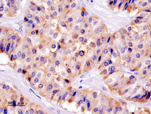
Insulinoma is the most common functional pancreatic neuroendocrine tumor. It is typically solitary, small (<2 cm), and benign (~90%), distributed fairly evenly through the pancreas, and it secretes insulin autonomously — causing fasting (and sometimes postprandial) hypoglycemia. Roughly 5–10% are multiple or malignant; multiplicity or a young patient should trigger evaluation for MEN1 (parathyroid → pituitary → pancreas).
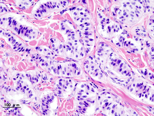
Draw the full panel during a hypoglycemic episode, when plasma glucose <55 mg/dL — values are meaningless when glucose is normal.
- Inappropriately high insulin ≥3 µU/mL — insulin should be suppressed when glucose is low
- C-peptide ≥0.6 ng/mL and proinsulin ≥5 pmol/L → confirms an endogenous β-cell source
- β-hydroxybutyrate ≤2.7 mmol/L — insulin suppresses ketogenesis, so hyperinsulinism = low ketones
- Glucose rises ≥25 mg/dL after 1 mg IV glucagon — insulin action has preserved hepatic glycogen
- Negative sulfonylurea/meglitinide screen and negative insulin antibodies
- 72-hour supervised fast is the gold-standard provocative test; terminate it once Whipple's triad + glucose <55 are met, then draw the labs above
Differentiating hyperinsulinemic (and non-insulin) states
| Condition | Insulin | C-peptide | Proinsulin | SU screen | β-OHB (ketones) |
|---|---|---|---|---|---|
| Insulinoma | High | High | High | Neg | Low |
| Sulfonylurea / meglitinide | High | High | Nml–high | Positive | Low |
| Exogenous (factitious) insulin | High | Low | Low | Neg | Low |
| IGF-2 tumor (NICTH) | Low | Low | Low | Neg | Low |
| Fasting / alcohol / adrenal insuff. | Low | Low | Low | Neg | High |
A 34-year-old nurse has recurrent episodes of sweating, tremor, and confusion that resolve with orange juice. During a captured episode, glucose is 38 mg/dL with markedly elevated insulin but suppressed C-peptide (<0.2 ng/mL) and a negative sulfonylurea screen.
- Diagnosis: exogenous (factitious) insulin administration — high insulin with a suppressed C-peptide means the insulin did not come from her own β-cells.
- Next best step: confirm the low C-peptide/proinsulin, screen for insulin analogs/antibodies, and involve psychiatry — do not proceed to pancreatic imaging or surgery.
- Board contrast: high C-peptide + positive drug screen = surreptitious sulfonylurea; high C-peptide + negative screen = insulinoma.
- Confirm the biochemistry first, THEN localize — never image before endogenous hyperinsulinism is proven
- Pancreatic-protocol contrast CT or MRI is first-line cross-sectional imaging
- Endoscopic ultrasound (EUS) is highly sensitive for small (<1–2 cm) tumors, especially in the head/body
- Occult tumors: selective arterial calcium stimulation with hepatic venous sampling regionalizes the lesion
- GLP-1 receptor (exendin) PET/SPECT is very sensitive for benign insulinoma; Ga-68 DOTATATE (somatostatin-receptor) imaging is less reliable because insulinomas variably express SSTR-2
- Intraoperative ultrasound + palpation improves detection at surgery
Management
Definitive therapy is surgical. Laparoscopic enucleation of a solitary benign insulinoma is curative in ~90%; larger, deep, or malignant lesions may need partial pancreatectomy.
Acute hypoglycemia: give fast-acting oral carbohydrate if the patient is alert; IV dextrose (D50) or IM/IV glucagon if consciousness is impaired.
Medical control (inoperable, metastatic, or as a bridge): diazoxide is first-line — it opens β-cell K-ATP channels, hyperpolarizing the cell to inhibit insulin release (side effects: fluid retention/edema, hirsutism; add a thiazide for the edema). Octreotide/lanreotide may help but can paradoxically worsen hypoglycemia by also suppressing glucagon.
Metastatic/malignant disease: everolimus (also directly raises glucose), sunitinib, and PRRT. Screen for MEN1 when tumors are multiple, the patient is young, or there is a suggestive family history.
A 28-year-old woman reports 6 months of early-morning and pre-lunch episodes of palpitations, confusion, and blurred vision relieved by eating; she has gained weight from constant snacking. A supervised 72-hour fast ends at 14 hours with glucose 41 mg/dL, insulin 12 µU/mL, C-peptide 1.4 ng/mL, elevated proinsulin, low β-OHB, and a negative sulfonylurea screen.
- Diagnosis: insulinoma — classic endogenous hyperinsulinemic hypoglycemia.
- Next best step: localize with pancreatic-protocol CT/MRI ± EUS — not more biochemistry.
- Definitive management: surgical enucleation (curative in the great majority of benign, solitary tumors).
- Whipple's triad (required to call true hypoglycemia): Symptoms of hypoglycemia → a Low measured plasma glucose → Relief when glucose is raised. All three are needed — a low meter reading without symptoms, or symptoms without a documented low glucose, does not qualify.
- C-peptide tells the source — "C = Came from me." High C-peptide = endogenous (insulinoma or sulfonylurea, split by the drug screen); low C-peptide with high insulin = exogenous injected insulin.
- Ketones split the causes: insulin and IGF-2 suppress ketogenesis → low β-OHB; alcohol, fasting, and adrenal insufficiency stay ketotic (high β-OHB). Alcohol classically blocks gluconeogenesis as NADH accumulates.
Practice Endocrine now
Board-style questions, spaced-repetition flashcards, and a Socratic AI tutor — free to start.