Hemodynamic Disorders: Edema, Thrombosis, Embolism & Infarction
A boards-focused walkthrough of the four core hemodynamic disorders — edema (Starling forces), thrombosis (Virchow's triad), embolism, and infarction — with classic vignette buzzwords, next-best-step decisions, and a red-vs-white infarct comparison.
The big picture
Hemodynamic disorders are derangements of fluid balance and blood flow, and Step 1 tests the mechanism behind each vignette rather than the label.
Edema is excess interstitial fluid, governed by Starling forces — net capillary movement set by hydrostatic pressure (pushes fluid out) and plasma oncotic/colloid pressure (holds fluid in). Thrombosis is pathologic clot formation inside an intact vessel, driven by Virchow's triad. A thrombus (or other material) that detaches and lodges downstream becomes an embolus, and vessel occlusion that starves tissue of perfusion produces an infarct.
These are mechanistically linked: a leg DVT (thrombosis) throws a pulmonary embolus that can cause a lung infarct. For every fact, ask: which Starling force shifted, which limb of Virchow's triad, and is the resulting infarct red or white?
- ↑ Hydrostatic pressure — CHF (generalized), DVT/venous obstruction (localized), portal hypertension
- ↓ Plasma oncotic pressure (hypoalbuminemia) — nephrotic syndrome, cirrhosis, protein-losing enteropathy, kwashiorkor
- Lymphatic obstruction → non-pitting lymphedema — filariasis (Wuchereria bancrofti), post-mastectomy/axillary dissection, tumor invasion
- ↑ Vascular permeability — inflammation, burns, sepsis, anaphylaxis → protein-rich leak
- Na⁺ / water retention — renal failure, RAAS activation
Transudate vs exudate (know the cutoffs):
- Transudate — protein-poor, specific gravity <1.012, clear; from hydrostatic/oncotic causes
- Exudate — protein-rich, SG >1.020, cellular/cloudy; from inflammation, infection, malignancy (Light's criteria separate the two in pleural effusions)
The three drivers of thrombosis — a bad wall, bad flow, or bad blood:
- Endothelial injury (wall) — atherosclerosis, vasculitis, HTN, smoking, hyperhomocysteinemia; dominant in arterial & cardiac thrombi
- Stasis / turbulent flow (flow) — immobilization, atrial fibrillation, aneurysm, hyperviscosity; dominant in venous thrombi
- Hypercoagulability (blood) — Factor V Leiden, prothrombin G20210A, protein C/S or antithrombin deficiency, OCPs/pregnancy, malignancy (Trousseau), antiphospholipid syndrome
Any single limb can nucleate a clot; postoperative and post-flight DVTs are the stasis limb in action.
- Lines of Zahn — alternating pale (platelet/fibrin) and dark (RBC) layers; signify an antemortem thrombus formed in flowing blood (absent in postmortem clots)
- Arterial / white thrombi — platelet-rich, form at endothelial injury/turbulence (over a plaque)
- Venous / red thrombi — RBC-rich, form in stasis (deep leg veins)
- Factor V Leiden — most common inherited hypercoagulable state; mutant factor V resists cleavage by activated protein C
- Protein C or S deficiency → warfarin-induced skin necrosis on warfarin initiation
- Antithrombin deficiency → blunted PTT response to heparin (heparin acts via antithrombin)
- Antiphospholipid syndrome → paradoxically prolonged PTT in vitro but thrombosis in vivo; recurrent miscarriage, false-positive VDRL/RPR
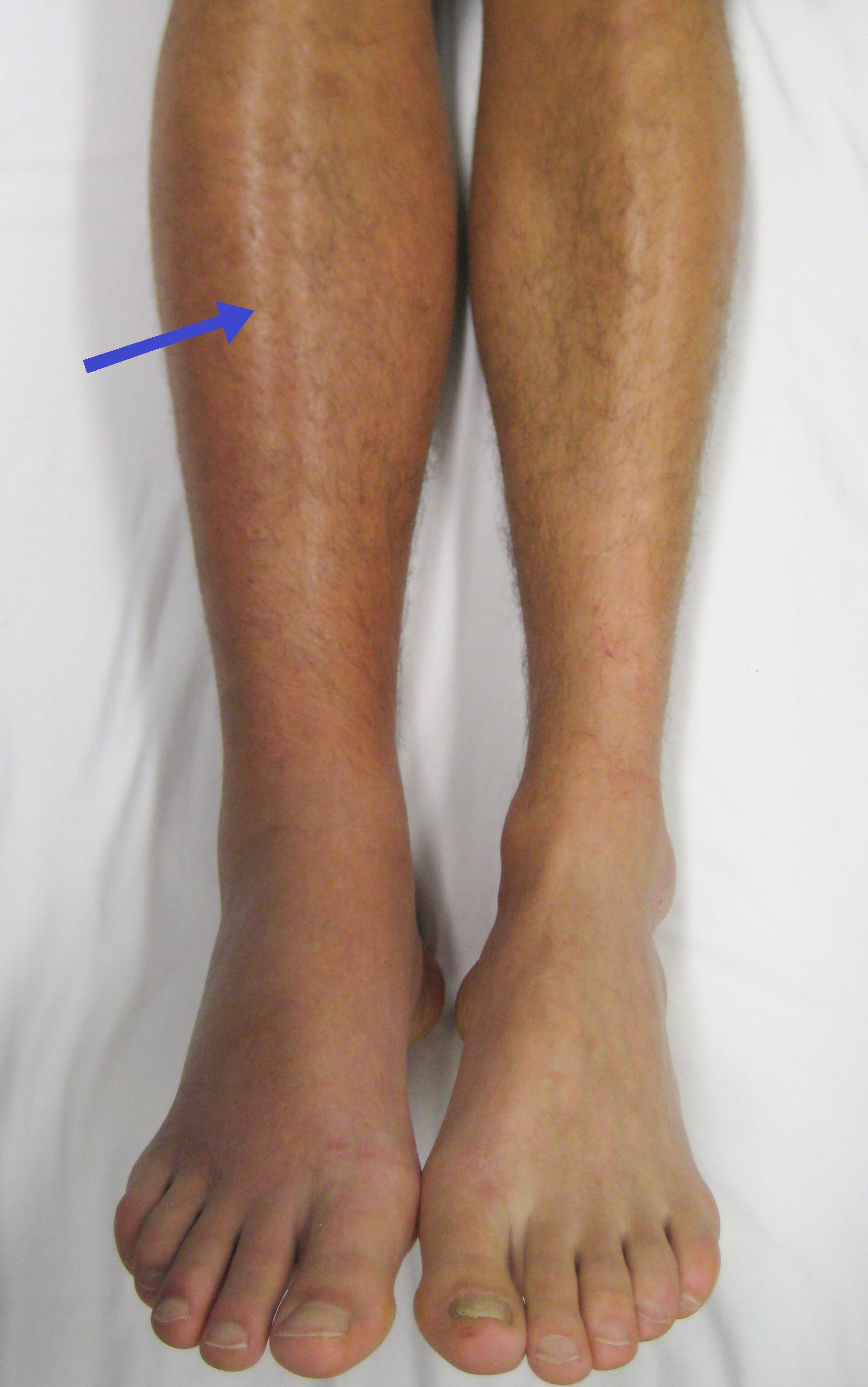
A 58-year-old woman develops sudden dyspnea and pleuritic chest pain 4 days after total knee replacement. HR 118, RR 28, SpO₂ 88%. Right calf is swollen and tender; ECG shows sinus tachycardia with a subtle S1Q3T3.
Diagnosis: Pulmonary embolism from a lower-extremity DVT (proximal deep veins — popliteal, femoral, iliac). Postoperative immobilization = the stasis limb of Virchow's triad.
Next best step:
- Hemodynamically stable → confirm with CT pulmonary angiography (Wells score guides testing; D-dimer only rules out PE in low-probability patients), then anticoagulate
- Hypotensive / unstable (massive PE) → thrombolysis
Sinus tachycardia is the most common ECG finding; large saddle emboli at the bifurcation can cause acute right-heart failure and sudden death.
A 22-year-old man sustains a femur fracture in a crash. ~48 hours later he becomes confused and dyspneic and develops a petechial rash over the chest and conjunctivae; SpO₂ 85%.
Diagnosis: Fat embolism syndrome — classic triad of hypoxia, neurologic changes, and petechiae, typically 24–72 h after long-bone/pelvic fracture or orthopedic surgery.
Next best step: Largely supportive (oxygen, ventilatory support); the diagnosis is clinical.
Contrast the other emboli tested:
- Amniotic fluid embolism — labor/peripartum → dyspnea, cardiovascular collapse, DIC
- Air embolism — decompression sickness ("the bends"), diving; procedures/central lines
- Paradoxical embolism — venous embolus reaches systemic arteries via a PFO/ASD/VSD
- Septic emboli — infected valve vegetations (infective endocarditis) shower to brain/kidney → abscesses, mycotic aneurysms
- Cholesterol (atheroembolism) — after cardiac cath/aortic manipulation → blue toes, cholesterol clefts, eosinophilia
- Coagulative necrosis in most infarcts; liquefactive necrosis in the brain
- Infarcts are typically wedge-shaped, with the occluded vessel at the apex and the organ surface at the base
- Vulnerability to hypoxia: neurons 3–5 min < myocardium 20–30 min < fibroblasts (hours) — neurons die first
- Organs with dual blood supply resist infarction: lung (bronchial + pulmonary), liver (hepatic artery + portal vein)
- Watershed infarcts form at borders of arterial territories (splenic flexure; ACA–MCA boundary) during systemic hypotension
- Slowly progressive occlusion permits collateral growth (stable angina) whereas abrupt occlusion infarcts (MI)
Red vs. white infarcts
| Feature | Red (hemorrhagic) infarct | White (anemic) infarct |
|---|---|---|
| Occlusion | Venous, or arterial with reperfusion | Arterial (end-artery) |
| Tissue type | Loose, or dual/collateral supply | Solid organ, single supply |
| Classic sites | Lung, small bowel, testis, ovary, liver | Heart, spleen, kidney |
| Gross look | Red, blood-filled, hemorrhagic | Pale, sharply demarcated, wedge |
| Necrosis | Coagulative | Coagulative |

Practice Pathology now
Board-style questions, spaced-repetition flashcards, and a Socratic AI tutor — free to start.