Glycolysis & Gluconeogenesis
A high-yield Step 1 review of glycolysis and gluconeogenesis: rate-limiting enzymes, the four irreversible bypass reactions, reciprocal control by fructose-2,6-bisphosphate/insulin/glucagon, and the classic tested diseases (pyruvate kinase deficiency, Von Gierke).
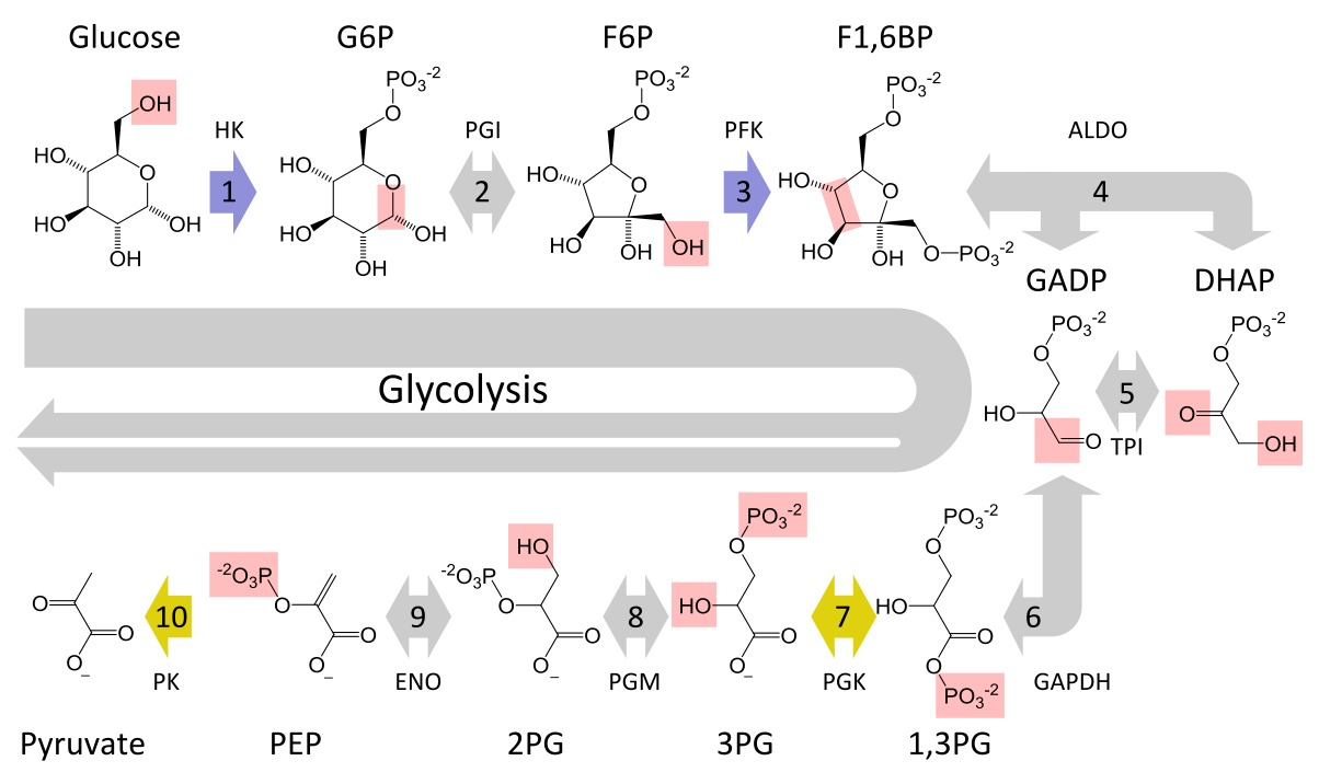
Glycolysis oxidizes glucose to 2 pyruvate in the cytosol of every cell, netting 2 ATP + 2 NADH — the sole ATP source for RBCs (no mitochondria). Gluconeogenesis runs the pathway in reverse, chiefly in the liver (also renal cortex, intestinal epithelium), to synthesize glucose during fasting from lactate, glucogenic amino acids (alanine), and glycerol. Three glycolytic steps are irreversible — hexokinase/glucokinase, PFK-1, pyruvate kinase — and gluconeogenesis bypasses each with a dedicated enzyme. The two pathways are reciprocally regulated so they never run at once (a wasteful futile cycle), coordinated by the fed/fasting hormones insulin and glucagon acting through fructose-2,6-bisphosphate. On the boards, expect questions on the rate-limiting enzymes, the required cofactors, and the classic deficiency diseases.
- Net yield: 2 ATP, 2 NADH, 2 pyruvate per glucose
- Rate-limiting enzyme: PFK-1 — activated by AMP and fructose-2,6-BP; inhibited by ATP and citrate
- Irreversible steps: hexokinase/glucokinase, PFK-1, pyruvate kinase (pyruvate kinase: PEP → pyruvate, substrate-level ATP)
- Purely glycolytic tissues (few/no mitochondria): RBCs, lens, cornea, renal medulla, leukocytes
- Arsenate substitutes for Pi at glyceraldehyde-3-phosphate dehydrogenase → glycolysis proceeds but yields no net ATP
- Arsenite (trivalent As) inhibits lipoic acid → blocks pyruvate dehydrogenase (and α-ketoglutarate dehydrogenase), so pyruvate can't enter the TCA cycle; vignette: garlic breath, watery/rice-water diarrhea, prolonged QT — treat with dimercaprol/succimer
- 4 irreversible bypass enzymes:
- Pyruvate carboxylase (mitochondria): pyruvate → oxaloacetate; needs biotin + ATP, allosterically activated by acetyl-CoA
- PEPCK (cytosol): oxaloacetate → PEP; needs GTP
- Fructose-1,6-bisphosphatase: rate-limiting; inhibited by AMP and F2,6-BP, activated by citrate
- Glucose-6-phosphatase (ER lumen; liver & kidney, also intestine): G6P → free glucose — muscle lacks it, so muscle cannot export glucose to blood
- Substrates: lactate, alanine, glycerol, propionyl-CoA (from odd-chain fatty acids)
- Even-chain fatty acids / acetyl-CoA CANNOT be converted to glucose — there is no acetyl-CoA → pyruvate reaction

Hexokinase vs. glucokinase
| Feature | Hexokinase | Glucokinase |
|---|---|---|
| Location | Most tissues | Liver, pancreatic β-cells |
| Km | Low (high affinity) | High (low affinity) |
| Vmax | Low | High |
| Induced by insulin | No | Yes |
| Inhibited by G6P | Yes | No |
| Role | Traps glucose even when scarce | Stores glucose when abundant; β-cell glucose sensor |
Vignette: A neonate is jaundiced with splenomegaly; an older sibling has had lifelong hemolytic anemia. Smear shows echinocytes (burr cells), no spherocytes and no Heinz bodies; Coombs negative, G6PD normal.
Dx: Pyruvate kinase deficiency (autosomal recessive) — the 2nd most common enzymatic cause of hereditary hemolytic anemia (after G6PD). RBCs cannot generate ATP → rigid membranes → extravascular hemolysis.
Key twist: upstream 2,3-BPG accumulates → right-shifted O2 dissociation curve → improved tissue O2 delivery, so the anemia is relatively well tolerated.
Next best step: confirm with a PK enzyme assay; management is supportive — folate, transfusions as needed, splenectomy in severe disease.
Vignette: An infant has a protuberant abdomen, doll-like facies, marked hepatomegaly, and seizures from fasting hypoglycemia. Labs: lactic acidosis, hyperuricemia (gout), hyperlipidemia; glucagon fails to raise blood glucose.
Dx: Von Gierke disease (glycogen storage disease type I) — glucose-6-phosphatase deficiency. Both glycogenolysis and gluconeogenesis generate G6P, but it cannot be dephosphorylated to free glucose → severe fasting hypoglycemia, and G6P is shunted to lactate, the pentose phosphate pathway (urate), and lipogenesis.
Next best step: frequent glucose/cornstarch feeds, avoid fructose and galactose, allopurinol for urate. Contrast with McArdle (type V) — muscle-only, exercise intolerance, no hypoglycemia.
"Pathway Produces Fresh Glucose" — the 4 bypass enzymes in pathway order (pyruvate → glucose):
- Pyruvate carboxylase (biotin; mitochondria)
- PEP carboxykinase / PEPCK (GTP)
- Fructose-1,6-bisphosphatase (rate-limiting)
- Glucose-6-phosphatase (ER; liver & kidney)
Biotin-dependent carboxylases to remember together: pyruvate carboxylase, acetyl-CoA carboxylase, propionyl-CoA carboxylase (all use biotin, CO2, and ATP).
The master switch: fructose-2,6-bisphosphate
Fructose-2,6-bisphosphate (F2,6-BP) decides which pathway runs: it activates PFK-1 (glycolysis) and inhibits fructose-1,6-bisphosphatase (gluconeogenesis). Its level is set by the bifunctional enzyme PFK-2/FBPase-2, switched by PKA phosphorylation:
- Fed / high insulin: enzyme is dephosphorylated → PFK-2 active → ↑F2,6-BP → glycolysis ON, gluconeogenesis off.
- Fasting / high glucagon (liver): ↑cAMP → PKA phosphorylates the enzyme → FBPase-2 active → ↓F2,6-BP → gluconeogenesis ON.
Reinforcing this, fasting β-oxidation raises acetyl-CoA, which activates pyruvate carboxylase (feeding gluconeogenesis) while inhibiting pyruvate dehydrogenase — the core biochemical logic of the fasting liver.
Practice Biochemistry now
Board-style questions, spaced-repetition flashcards, and a Socratic AI tutor — free to start.