GERD & Peptic Ulcer Disease
A boards-focused walk through GERD and peptic ulcer disease — from LES failure and the acid-versus-mucosa imbalance to the classic duodenal-vs-gastric ulcer split, H. pylori testing rules, the dyspepsia next-best-step algorithm, eradication regimens, and the perforation/bleed/Zollinger-Ellison complications examiners love.
Acid vs Mucosa: One Theme, Two Diseases
Both GERD and peptic ulcer disease (PUD) come down to acid overwhelming a mucosal barrier — the difference is where.
GERD = reflux of gastric contents into the esophagus, driven by a failing lower esophageal sphincter (LES): transient LES relaxations (the dominant mechanism), low resting tone, a hiatal hernia, or delayed gastric emptying. Chronic acid injury to squamous mucosa → erosive esophagitis, peptic stricture, and Barrett esophagus — metaplastic columnar epithelium with goblet cells (intestinal metaplasia) that is the precursor to esophageal adenocarcinoma.
PUD = a mucosal break (≥5 mm, through the muscularis mucosae) in the stomach or duodenum, from an imbalance of aggressive factors (acid, pepsin, H. pylori, NSAIDs) versus protective ones (mucus, bicarbonate, prostaglandins, mucosal blood flow). Two causes dominate:
- *H. pylori* — a urease-producing, curved gram-negative rod; classically behind ~90% of duodenal and up to ~70% of gastric ulcers.
- NSAIDs — block COX → ↓ mucosal prostaglandins; the classic driver of gastric ulcers.
Rare but heavily tested: Zollinger–Ellison syndrome (gastrinoma).
- *H. pylori* is urease-positive, spreads fecal–oral, and colonizes the gastric antrum → ↓ somatostatin → ↑ gastrin → ↑ acid; it is a class I (IARC group 1) carcinogen → gastric adenocarcinoma + MALT lymphoma (which can regress with eradication)
- Duodenal ulcer pain is RELIEVED by food (classically wakes the patient at night, ~2–3 h postprandial); gastric ulcer pain is WORSE with food → food avoidance and weight loss
- Every gastric ulcer is biopsied to exclude malignancy, and is typically re-scoped in 8–12 weeks to confirm healing; duodenal ulcers are essentially never malignant
- PPIs > H2 blockers and heal faster: ~8 weeks for erosive GERD and gastric ulcers, ~4 weeks for duodenal ulcers
- Hold the PPI ~2 weeks before urea breath test, stool antigen, or biopsy urease test — PPIs cause false negatives
- Serology (IgG) can't distinguish active from past infection — never use it to confirm cure
- Confirm eradication with urea breath test or stool antigen ≥4 weeks after finishing therapy
- Anterior duodenal ulcer → perforation (free air); posterior → gastroduodenal artery bleed
Duodenal vs Gastric Ulcer
| Feature | Duodenal ulcer | Gastric ulcer |
|---|---|---|
| Pain & food | Relieved by food; pain ~2–3 h later & at night | Worse with food → food fear, weight loss |
| H. pylori | ~90% | ~70% (NSAIDs a major cause) |
| Acid output | Normal–high | Normal or low |
| Malignant potential | Essentially never | Can be malignant → biopsy |
| Classic site | 1st part of duodenum | Lesser curvature / antrum |
| Bleeding vessel | Posterior → gastroduodenal artery | Lesser curve → left gastric artery |
| Typical age | Younger | Older |
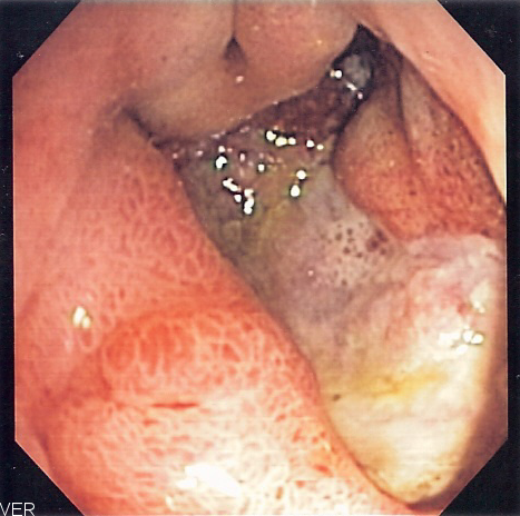
ALARM — red-flag features in dyspepsia/GERD that mandate prompt upper endoscopy (EGD) instead of an empiric acid-suppression trial:
- A — Anemia (iron-deficiency) / Anorexia
- L — Loss of weight (unintentional)
- A — Abdominal mass or new-onset dysphagia
- R — Recent-onset, progressive symptoms
- M — Melena / hematemesis (GI bleeding)
Also scope for odynophagia or a family history of upper-GI cancer, and for new dyspepsia at age ≥60 (per ACG, regardless of alarm features). No alarm features + age <60 → test-and-treat for H. pylori or trial a PPI first.
- 42-y/o, epigastric burning, no alarm features: don't scope first — test-and-treat. Check H. pylori by urea breath test or stool antigen; if positive, eradicate (if negative, empiric PPI trial).
- 68-y/o, new dyspepsia + 5-kg weight loss + iron-deficiency anemia: EGD now — age ≥60 and alarm features.
- Typical heartburn/regurgitation, no alarm features: empiric PPI trial × 8 weeks — no EGD needed initially.
- EGD shows a gastric ulcer: biopsy the margins (exclude malignancy) and repeat EGD in 8–12 weeks to confirm healing.
- Heartburn persists on high-dose PPI with a normal EGD: confirm pathologic reflux with ambulatory pH (or pH-impedance) monitoring; add esophageal manometry before any antireflux surgery (to exclude achalasia/dysmotility).
Management: PPIs and the Eradication Regimens
GERD. Begin lifestyle measures (weight loss, elevate the head of the bed, avoid late/large meals, trigger foods, and tobacco) plus a PPI (~8 weeks) — more effective than H2 blockers — then step down to the lowest effective dose. Refractory disease → confirm pathologic reflux with pH monitoring; durable options include Nissen fundoplication (also for large hiatal hernia or PPI intolerance). Barrett esophagus → surveillance EGD; dysplasia → endoscopic ablation/resection.
PUD. Stop NSAIDs, start a PPI, and eradicate *H. pylori* when present:
- Bismuth quadruple therapy — PPI + bismuth + tetracycline + metronidazole × 14 days. Preferred first-line, especially with prior macrolide use or high local clarithromycin resistance.
- Clarithromycin triple therapy — PPI + clarithromycin + amoxicillin × 14 days — only where clarithromycin resistance is <15% and there's no prior macrolide exposure.
Always confirm eradication (urea breath test / stool antigen ≥4 weeks later). An NSAID-dependent patient needs a co-prescribed PPI for prophylaxis.
H. pylori Testing at a Glance
| Test | Detects | Board pearls |
|---|---|---|
| Urea breath test | Active infection | Confirms cure; hold PPI ~2 wk (false −) |
| Stool antigen | Active infection | Confirms cure; inexpensive; hold PPI |
| Rapid urease / histology | Active (biopsy at EGD) | PPI → false negative; histology also shows gastritis |
| Serology (IgG) | Past or present | Can't confirm eradication; not affected by PPI |

- Sudden severe epigastric pain, rigid "board-like" abdomen, free air under the diaphragm on upright CXR: perforated ulcer (classically an anterior duodenal ulcer) → resuscitate + emergency surgery.
- Hematemesis/melena with hypotension: think a posterior duodenal ulcer eroding the gastroduodenal artery → IV fluids/transfuse, IV PPI, urgent EGD for diagnosis and hemostasis.
- Early satiety, vomiting of undigested food, a succussion splash: gastric outlet obstruction.
- Multiple or refractory ulcers, ulcers distal to the duodenal bulb, diarrhea (± MEN1): Zollinger–Ellison syndrome → fasting serum gastrin (markedly ↑), then a secretin stimulation test (paradoxical rise in gastrin). Hold the PPI first — it raises gastrin and causes false positives.
Practice GI now
Board-style questions, spaced-repetition flashcards, and a Socratic AI tutor — free to start.