Gallstones & Biliary Disease
A boards-focused walkthrough of biliary disease from lithogenic bile to next-best-step management, mapping each syndrome (colic, cholecystitis, choledocholithiasis, cholangitis, gallstone pancreatitis) to its buzzwords, labs, imaging, and definitive intervention. Emphasizes ultrasound-first workup, HIDA for equivocal cholecystitis, and ERCP for CBD stones and cholangitis.
Pathophysiology: lithogenic bile and where the stone lodges
Gallstones (cholelithiasis) form when bile turns lithogenic. Cholesterol stones (the majority — ~80–90% in the US) require three ingredients: cholesterol supersaturation of bile, gallbladder hypomotility/stasis, and accelerated nucleation. Pigment stones are calcium bilirubinate — black stones form in chronic hemolysis (sickle cell, hereditary spherocytosis, thalassemia) and cirrhosis; brown stones form within the ducts under stasis/infection (bacterial β-glucuronidase, Asian liver flukes such as Clonorchis).
The clinical syndrome reflects where a stone impacts:
- Intermittent cystic-duct obstruction → biliary colic.
- Sustained cystic-duct obstruction + inflammation → acute cholecystitis.
- Passage into the common bile duct (CBD) → choledocholithiasis, which can progress to ascending cholangitis (infection behind an obstruction) or, lodging at the ampulla, gallstone pancreatitis.
Key board point: most gallstones are asymptomatic and need no treatment.
- Cholesterol-stone risk — the 5 F's: Female, Forty, Fat, Fertile, Fair. Add estrogen/OCPs, rapid weight loss or bariatric surgery, prolonged TPN, and fibrates.
- Ileal disease/resection (Crohn's) → bile-salt malabsorption → cholesterol stones. Native American (Pima) ethnicity has the highest prevalence.
- Black pigment stones: chronic hemolysis (sickle cell, spherocytosis, thalassemia) and cirrhosis.
- Brown pigment stones: biliary infection/stasis, liver flukes (Clonorchis sinensis); typically intraductal.
- X-ray visibility: only ~10–15% of stones are radiopaque (calcium); most cholesterol stones are radiolucent → ultrasound, not plain film, is the test.
- Prophylactic cholecystectomy for asymptomatic stones is reserved for a porcelain gallbladder, stones >3 cm, or an anomalous pancreaticobiliary junction.
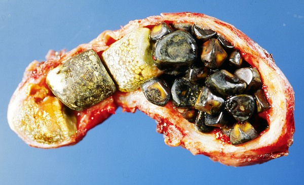
Comparison of biliary syndromes
| Condition | Pain & systemic signs | Labs | Imaging / dx | Next step |
|---|---|---|---|---|
| Biliary colic | RUQ pain <6 h after fatty meal, no fever | Normal | US: stones, no wall change | Elective lap chole |
| Acute cholecystitis | RUQ pain >6 h, fever, +Murphy sign | ↑WBC, mild ↑LFTs | US: wall >3 mm, pericholecystic fluid; HIDA if equivocal | IVF, antibiotics, early lap chole |
| Choledocholithiasis | RUQ pain + jaundice | ↑ALP/GGT, ↑direct bili, CBD >6 mm | US → MRCP/EUS; ERCP if high-risk | ERCP extraction, then chole |
| Ascending cholangitis | Charcot triad ± Reynolds pentad | ↑ALP/bili, ↑WBC, +cultures | Dilated CBD + stone | IVF, abx, urgent ERCP decompression |
| Gallstone pancreatitis | Epigastric pain radiating to back | ↑lipase, ALT >3× ULN | US: stones ± dilated CBD | Supportive; ERCP if cholangitis/obstruction; chole same admission |
Vignette: A 45-year-old obese woman has 8 hours of constant RUQ pain, fever 38.4°C, and nausea after a fatty meal. Palpation of the RUQ during inspiration causes her to catch her breath. WBC 14,000, mildly elevated ALT/AST, normal bilirubin. RUQ ultrasound shows gallstones but no wall thickening and an equivocal sonographic Murphy sign.
- Diagnosis: Acute cholecystitis (positive Murphy sign + fever + leukocytosis).
- Best next step when US is equivocal: HIDA scan (cholescintigraphy) — non-visualization of the gallbladder confirms cystic-duct obstruction (highest sensitivity/specificity).
- Management: NPO, IV fluids, analgesia, IV antibiotics, and early laparoscopic cholecystectomy (within ~72 h). If the patient is too unstable or a poor surgical candidate → percutaneous cholecystostomy as a bridge.

- 5 F's — cholesterol-stone risk: Female, Forty, Fat, Fertile, Fair.
- Charcot triad — ascending cholangitis: fever, jaundice, RUQ pain.
- Reynolds pentad — suppurative cholangitis: Charcot triad + hypotension + altered mental status → emergent biliary decompression.
- Rigler triad — gallstone ileus: pneumobilia + small-bowel obstruction + ectopic (RLQ) gallstone on imaging.
- First test for any RUQ biliary complaint: transabdominal ultrasound.
- Acute cholecystitis on US: stones, wall thickening >3 mm, pericholecystic fluid, sonographic Murphy. Equivocal → HIDA (non-visualization = positive).
- CBD-stone (choledocholithiasis) high-risk predictors (2019 ASGE): CBD stone seen on imaging, total bilirubin >4 mg/dL with a dilated CBD, or clinical cholangitis → go straight to ERCP.
- Intermediate risk (abnormal LFTs, age >55, or dilated CBD alone): confirm first with MRCP or EUS before committing to ERCP.
- ERCP is therapeutic (sphincterotomy + stone extraction) and carries a post-ERCP pancreatitis risk — avoid using it purely diagnostically when MRCP/EUS suffices.
- Cholestatic pattern (↑ALP, ↑GGT, ↑direct bilirubin) = ductal obstruction; a hepatocellular bump with ALT >3× ULN points to gallstone pancreatitis.
Vignette: A 70-year-old man has RUQ pain, fever with rigors, and jaundice. BP 88/54, and he is confused. WBC 18,000, total bilirubin 6 mg/dL, ↑ALP; ultrasound shows a dilated common bile duct with a stone.
- Diagnosis: Ascending (acute) cholangitis — Charcot triad plus hypotension and altered mentation = Reynolds pentad (suppurative).
- Immediate management: aggressive IV fluids and broad-spectrum IV antibiotics covering enteric gram-negatives/anaerobes; ICU-level resuscitation.
- Definitive next step: urgent/emergent biliary decompression by ERCP (sphincterotomy + stone removal). If ERCP fails or is unavailable → percutaneous transhepatic cholangiography (PTC) drainage.
- Cholecystectomy is done after the patient stabilizes, not acutely.
- Acalculous cholecystitis: critically ill/ICU, TPN, major trauma, burns, sepsis; no stones, high mortality → often percutaneous cholecystostomy first.
- Porcelain gallbladder: calcified wall on imaging; associated with gallbladder adenocarcinoma → cholecystectomy.
- Gallstone ileus: cholecystoenteric fistula lets a large stone impact at the ileocecal valve → SBO; look for Rigler triad.
- Mirizzi syndrome: a stone in the cystic duct/gallbladder neck externally compresses the common hepatic duct → obstructive jaundice.
- Gallbladder cancer: risk factors — porcelain gallbladder, large stones (>3 cm), chronic cholecystitis; usually presents late.
- Post-cholecystectomy recurrent RUQ pain/jaundice → suspect retained CBD stone or bile leak → MRCP/ERCP.
Practice GI now
Board-style questions, spaced-repetition flashcards, and a Socratic AI tutor — free to start.