Diabetes Mellitus & Diabetic Ketoacidosis
A boards-focused review of diabetes mellitus — separating type 1 from type 2, the diagnostic thresholds, and chronic complications — then a deep dive on diabetic ketoacidosis: pathophysiology, the classic vignette, the potassium trap, the ketone-testing pitfall, and DKA versus HHS.
The two diseases behind one word
Diabetes mellitus (DM) is a family of disorders united by chronic hyperglycemia arising from defective insulin secretion, insulin action, or both. Two forms dominate the boards. Type 1 DM is a T-cell–mediated autoimmune destruction of pancreatic β-cells, producing an absolute insulin deficiency in a classically young, lean patient. Type 2 DM combines peripheral insulin resistance with a progressive β-cell secretory defect (relative deficiency) and is tightly linked to obesity and the metabolic syndrome.
Sustained hyperglycemia injures tissue by two mechanisms worth memorizing: nonenzymatic glycation (advanced glycation end-products damaging vessel walls and basement membranes) and the polyol/sorbitol pathway, in which aldose reductase traps sorbitol inside cells that have little or no sorbitol dehydrogenase — lens, retina, Schwann cells, and kidney — driving osmotic and oxidative injury. Together these produce the micro- and macrovascular complications responsible for most diabetic morbidity.
Type 1 vs Type 2 DM
| Feature | Type 1 DM | Type 2 DM |
|---|---|---|
| Typical patient | Child/adolescent, lean | Adult, obese (increasingly younger) |
| Core defect | Autoimmune β-cell loss → absolute deficiency | Insulin resistance + relative deficiency |
| Autoantibodies | Anti-GAD65, anti-islet cell, anti-insulin | Absent |
| Genetics | HLA-DR3/DR4; ~30–50% twin concordance | Polygenic; ~90% twin concordance (stronger) |
| C-peptide | Low / undetectable | Normal or high (early) |
| Islet histology | Insulitis (lymphocytic infiltrate) | Amyloid (amylin/IAPP) deposits |
| Acute crisis | DKA | HHS (hyperosmolar) |
| First-line therapy | Insulin (required for life) | Lifestyle + metformin, then add-ons |
- Diagnostic criteria (any one; confirm on a repeat test unless unequivocally symptomatic): HbA1c ≥ 6.5%, fasting plasma glucose ≥ 126 mg/dL, 2-hr 75-g OGTT ≥ 200 mg/dL, or random glucose ≥ 200 mg/dL with classic symptoms.
- Prediabetes: HbA1c 5.7–6.4%, FPG 100–125, OGTT 140–199.
- HbA1c reflects ~3-month glycemia; falsely low with shortened RBC lifespan (hemolytic anemia, recent bleeding/transfusion) and falsely high with iron-deficiency anemia (older, long-lived RBCs).
- Microvascular (tracks glycemic control): retinopathy; nephropathy — Kimmelstiel–Wilson nodular glomerulosclerosis, earliest sign is moderately increased albuminuria (microalbuminuria); neuropathy — stocking-glove sensory loss.
- Macrovascular: coronary artery disease (leading cause of death), stroke, peripheral arterial disease.
- Renal/cardiac protection: start an ACE inhibitor or ARB once albuminuria appears; add an SGLT2 inhibitor to slow CKD progression and cut heart-failure/CV events (now standard cardiorenal therapy).
How DKA happens
Diabetic ketoacidosis ignites when profound insulin deficiency is amplified by a surge of counter-regulatory hormones — glucagon, catecholamines, cortisol, and growth hormone. The low-insulin/high-glucagon state does two destructive things at once:
- Unrestrained hepatic gluconeogenesis and glycogenolysis → hyperglycemia → osmotic diuresis with volume and electrolyte loss.
- Adipose lipolysis floods the liver with free fatty acids that undergo β-oxidation into ketoacids — β-hydroxybutyrate (predominant) and acetoacetate.
Ketoacid accumulation generates a high anion-gap metabolic acidosis. DKA is the classic crisis of type 1 DM; the most common precipitant is infection, followed by insulin nonadherence and new-onset disease.

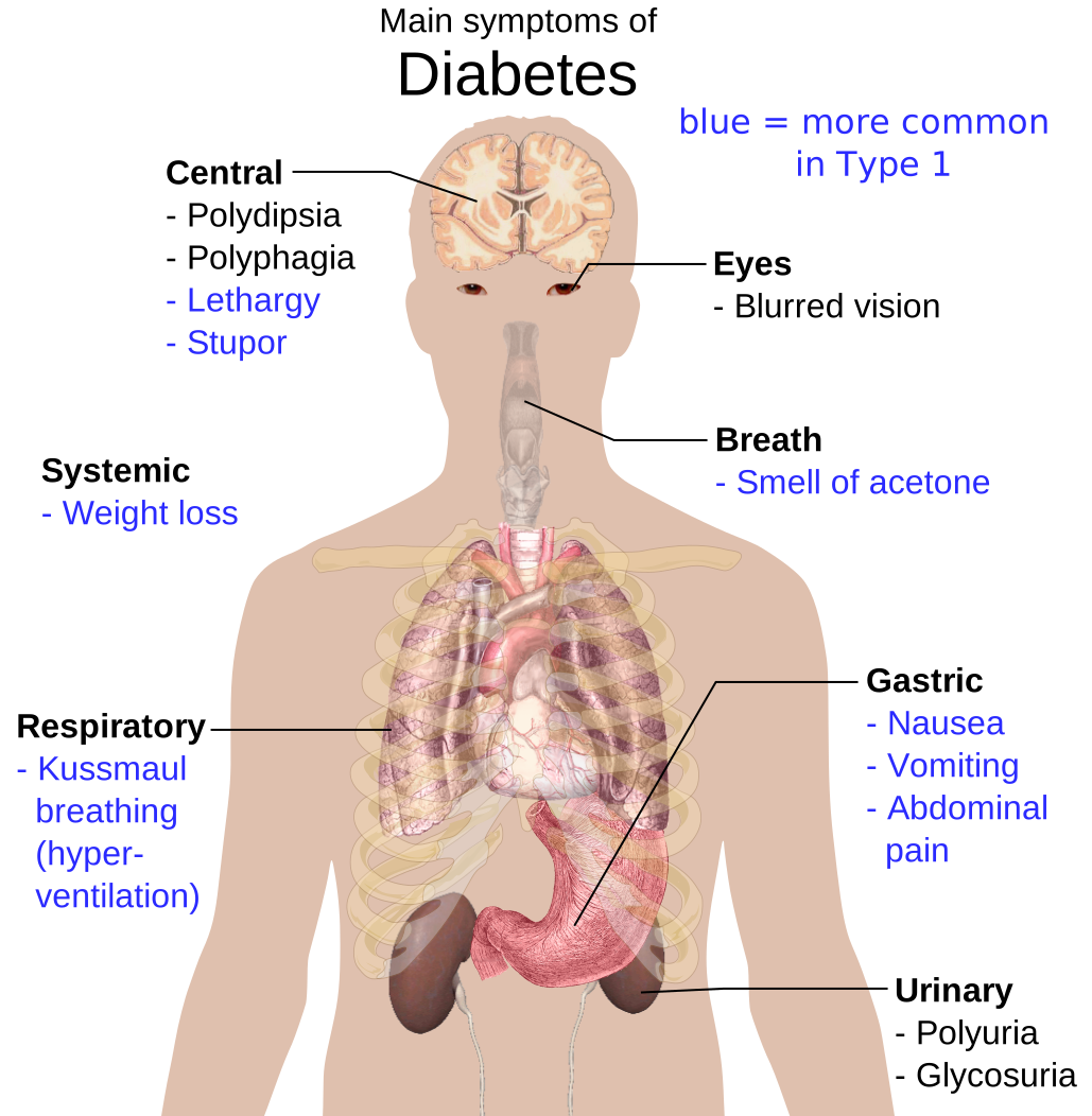
Buzzwords: a young type 1 diabetic (or first presentation) with polyuria, polydipsia, nausea/vomiting, and abdominal pain, breathing in deep, rapid Kussmaul respirations with a fruity (acetone) breath odor and signs of dehydration. Labs: glucose ~250–600, pH < 7.3, HCO3 < 18, positive serum/urine ketones, and an elevated anion gap.
Management, in order:
- IV isotonic fluids (0.9% saline) — the first and single most important step.
- IV regular insulin — after checking potassium.
- Potassium repletion — total-body K is depleted; insulin will drive K into cells. If K < 3.3 mEq/L, hold insulin and give potassium first.
- Add dextrose once glucose reaches ~200 mg/dL, and continue insulin until the anion gap closes — the true endpoint, not mere glucose normalization.
The tested trap: presenting serum potassium is often normal or high despite whole-body depletion, because acidosis and insulin lack shift K out of cells — then it plummets with treatment.
- Anion gap = Na − (Cl + HCO3); widened by unmeasured ketoanions.
- Potassium pitfall: serum K is often normal/↑ at presentation despite total-body depletion — check K before insulin, replete early, and monitor closely, because insulin drives a sharp intracellular shift. Hold insulin if K < 3.3 mEq/L until repleted.
- Corrected sodium: add ~1.6 mEq/L to measured Na for every 100 mg/dL glucose above 100. Hyperglycemia osmotically pulls water into the blood, causing a true dilutional (hypertonic) hyponatremia — not the lab-artifact 'pseudohyponatremia' of hyperlipidemia.
- Fluids come before insulin.
- Treat until the anion gap normalizes (not just glucose); overlap subcutaneous insulin with the IV drip for ~1–2 hr before stopping it to prevent rebound ketosis.
- Ketone-testing trap: the nitroprusside (urine/serum) assay detects acetoacetate, not β-hydroxybutyrate — so measured ketones can paradoxically rise early in therapy as β-hydroxybutyrate converts to acetoacetate; track the anion gap or direct β-hydroxybutyrate instead.
- Cerebral edema is the most feared treatment complication, especially in children — avoid overly rapid correction of glucose/osmolality.
- Euglycemic DKA: suspect with SGLT2 inhibitors, pregnancy, or starvation — acidosis and ketones with near-normal glucose.
DKA vs HHS
| Feature | DKA | HHS |
|---|---|---|
| Typical patient | Type 1 DM | Type 2 DM, elderly |
| Glucose | > 250 (usually 250–600) | > 600 (often > 1000) |
| Ketones | Marked | Minimal / absent |
| Arterial pH | < 7.3 (acidotic) | > 7.3 (normal) |
| Serum HCO3 | < 18 | > 18 (normal) |
| Anion gap | High | Normal / mildly ↑ |
| Serum osmolality | Variable | Very high (> 320 mOsm/kg) |
| Mental status | Alert → drowsy | Marked obtundation / coma |
| Mortality | Lower (~1–5%) | Higher (~10–20%) |
The classic triggers of DKA all start with I:
- Infection — the most common precipitant
- Infarction — MI or stroke (counter-regulatory surge)
- Insulin lack — nonadherence or new-onset type 1 DM
- Indiscretion — dietary noncompliance
- Infant — pregnancy
Search for the trigger while you treat: any DKA warrants a hunt for infection or ischemia.
Practice Endocrine now
Board-style questions, spaced-repetition flashcards, and a Socratic AI tutor — free to start.