Cushing Syndrome & Adrenal Insufficiency
A boards-focused walkthrough of Cushing syndrome and adrenal insufficiency: HPA-axis pathophysiology, the confirm-then-localize diagnostic sequence with dynamic testing, and next-best-step management (including adrenal crisis), anchored by classic vignette buzzwords and lab-comparison tables.
Cortisol & the HPA axis
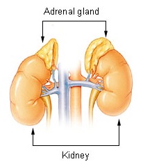
Cortisol is made in the adrenal cortex (zona fasciculata) under drive from pituitary ACTH, itself driven by hypothalamic CRH; cortisol exerts negative feedback on both. Cushing syndrome = chronic cortisol EXCESS; adrenal insufficiency (AI) = cortisol DEFICIENCY.
Physiology the boards exploit:
- Aldosterone (zona glomerulosa) is governed by RAAS and potassium, largely ACTH-independent — so it is preserved in secondary AI but lost in primary AI.
- Cortisol is catabolic (muscle, skin, bone), anti-insulin (gluconeogenesis → hyperglycemia), immunosuppressive, and permissive for catecholamine vascular tone (↑α₁ receptors) — losing it causes refractory hypotension.
- ACTH is cleaved from POMC, which also yields MSH; therefore high-ACTH states cause hyperpigmentation.
Causes
- Most common overall: exogenous (iatrogenic) glucocorticoids.
- ACTH-dependent (endogenous): Cushing disease = pituitary ACTH-secreting adenoma (most common endogenous cause, ~70%); ectopic ACTH (small cell lung cancer, bronchial carcinoid).
- ACTH-independent: adrenal adenoma, carcinoma, or bilateral hyperplasia → ACTH suppressed.
Presentation (vignette buzzwords)
- Central obesity, moon facies, dorsocervical fat pad ("buffalo hump"), wide (>1 cm) violaceous abdominal striae.
- Proximal muscle weakness, thin skin, easy bruising.
- Hypertension, hyperglycemia/new diabetes, osteoporosis, infections, mood change/psychosis.
- Rapid onset + weight loss + severe hypokalemic metabolic alkalosis + marked hyperpigmentation → ectopic ACTH (paraneoplastic).
Cushing: diagnostic sequence
Work in two phases — first prove hypercortisolism, then localize.
1. Confirm hypercortisolism (exclude exogenous steroids by history first; obtain ≥2 abnormal screening tests):
- 1-mg overnight low-dose dexamethasone suppression — normally cortisol falls <1.8 µg/dL; failure to suppress is positive.
- Late-night salivary cortisol (loss of the normal nighttime nadir).
- 24-hour urinary free cortisol.
2. Localize with plasma ACTH:
- Low/suppressed ACTH → ACTH-independent → adrenal CT.
- Normal/high ACTH → ACTH-dependent → distinguish pituitary vs ectopic using high-dose dexamethasone (pituitary suppresses, ectopic does not), CRH stimulation (pituitary responds), pituitary MRI, and inferior petrosal sinus sampling (IPSS) — the gold standard when MRI is equivocal.

Vignette: A 40-year-old woman has 6 months of central weight gain, facial rounding, purple abdominal striae, proximal weakness, easy bruising, and new hypertension with hyperglycemia.
Dx: Clinical suspicion of Cushing syndrome.
Next best step: Confirm biochemically first — do NOT image first. Order a screening test for hypercortisolism (1-mg overnight dexamethasone suppression, late-night salivary cortisol, or 24-h urinary free cortisol). Only after biochemical confirmation do you measure plasma ACTH, then obtain targeted imaging.
Board trap: Jumping straight to pituitary MRI — nonfunctioning pituitary incidentalomas are common and will mislead you.
ACTH-dependent vs -independent Cushing
| Feature | Cushing disease (pituitary) | Ectopic ACTH | Adrenal tumor |
|---|---|---|---|
| Plasma ACTH | High–normal | Very high | Low / suppressed |
| High-dose dexamethasone | Suppresses (>50%) | No suppression | No suppression |
| CRH stimulation | ACTH/cortisol rise | No response | No response |
| Best imaging | Pituitary MRI | Chest/abdomen CT | Adrenal CT |
| Classic clue | Woman 20–40, slow onset | SCLC/carcinoid; ↓K⁺, rapid | Unilateral adrenal mass |
Primary (Addison disease) — destruction of the adrenal cortex → loss of cortisol + aldosterone + adrenal androgens. High ACTH (POMC) → hyperpigmentation.
- Causes: autoimmune adrenalitis (most common in developed world), TB (leading cause worldwide), adrenoleukodystrophy, metastases, bilateral hemorrhage (Waterhouse–Friderichsen, meningococcemia).
Secondary/tertiary — deficient pituitary ACTH or hypothalamic CRH. Most common cause overall = abrupt withdrawal of chronic glucocorticoids (suppressed HPA axis). Cortisol low, but aldosterone preserved (RAAS intact) → no hyperpigmentation, no hyperkalemia.
Labs/clues: fatigue, weight loss, hypotension, hyponatremia (both). Hyperkalemia + metabolic acidosis + salt craving → primary only. Hypoglycemia common.
Dx: 8 AM cortisol (low), then ACTH (high = primary; low/normal = secondary), confirm with the cosyntropin (ACTH) stimulation test.
Vignette: A patient with known Addison disease (or on chronic glucocorticoids) develops an intercurrent infection with vomiting, then hypotension refractory to IV fluids and pressors, abdominal pain, and hypoglycemia + hyponatremia. In primary AI expect hyperkalemia too (aldosterone loss); a secondary (steroid-withdrawal) crisis usually spares potassium.
Dx: Adrenal (Addisonian) crisis.
Next best step: Treat immediately — do not wait for labs. Give IV hydrocortisone 100 mg plus aggressive IV normal saline with dextrose, and treat the precipitant. Draw cortisol/ACTH but do not delay steroids (pressors fail without cortisol's permissive effect).
- If the diagnosis is uncertain and you want to preserve testing, give dexamethasone (does not cross-react with the cortisol assay) and then perform the cosyntropin stimulation test.
- Chronic Rx: hydrocortisone; add fludrocortisone for primary AI; stress-dose during illness/surgery; medical-alert ID.

Primary vs secondary AI — labs & axes
| Feature | Primary (Addison) | Secondary / Tertiary |
|---|---|---|
| Defect | Adrenal cortex | Pituitary ACTH / hypothalamic CRH |
| ACTH | High | Low / inappropriately normal |
| Cortisol | Low | Low |
| Aldosterone | Low | Normal (RAAS intact) |
| Hyperpigmentation | Yes | No |
| Serum K⁺ | High | Normal |
| Serum Na⁺ | Low | Low / normal |
| Cosyntropin stim | Blunted rise | May respond (if recent onset) |
| Mineralocorticoid Rx | Fludrocortisone needed | Not needed |
CUSHINGOID — signs of chronic glucocorticoid excess (endogenous or iatrogenic):
- C — Cataracts (posterior subcapsular)
- U — peptic Ulcers
- S — Striae / Skin thinning & easy bruising
- H — Hypertension / Hirsutism
- I — Infections (immunosuppression)
- N — avascular Necrosis (femoral head)
- G — Glucose intolerance → hyperglycemia/diabetes
- O — Osteoporosis / central Obesity
- I — Impaired wound healing
- D — Depression / mood change
AI memory hook: Addison = A darker patient (high ACTH/POMC → hyperpigmentation) with low Na⁺ / high K⁺. Secondary AI = pale, with normal K⁺ (aldosterone spared).
Practice Endocrine now
Board-style questions, spaced-repetition flashcards, and a Socratic AI tutor — free to start.