Cirrhosis & Portal Hypertension
End-stage liver fibrosis produces two linked problems — hepatocellular synthetic failure and portal hypertension — and nearly every board fact (varices, ascites, SBP, encephalopathy, hyperestrinism, Child-Pugh/MELD) flows from one of them. This lesson maps the causes, portosystemic collaterals, decompensating complications, and next-best-step management.
The two-problem framework
Cirrhosis is the final common pathway of chronic liver injury: diffuse bridging fibrosis plus regenerative nodules that destroy normal hepatic architecture. That architectural distortion creates exactly two downstream problems, and almost every testable finding maps onto one of them:
- Hepatocellular (synthetic) failure — the liver can no longer make what it should: ↓ albumin, ↓ clotting factors (↑ PT/INR), impaired estrogen and ammonia clearance, ↓ thrombopoietin.
- Portal hypertension — fibrosis and nodules ↑ intrahepatic resistance, backing pressure up into the portal system: varices, ascites, splenomegaly, and — via portosystemic shunting — hepatic encephalopathy.
Hepatic encephalopathy actually straddles both problems (shunting plus lost hepatocellular ammonia clearance), but for every other finding the reflex works: when a vignette drops a clue, ask "synthesis or pressure?" — it organizes the entire topic.
- Cirrhosis = bridging fibrosis + regenerative nodules; portal HTN defined by HVPG (hepatic venous pressure gradient): >5 = portal HTN, ≥10 = clinically significant (decompensation/varices), ≥12 = variceal bleeding threshold.
- Leading causes: chronic HCV, alcohol, and MASLD/NASH (formerly NAFLD — now rising fastest); also HBV, hemochromatosis, Wilson disease, α1-antitrypsin deficiency, PBC/PSC, autoimmune hepatitis.
- Synthetic failure labs: ↓ albumin, ↑ PT/INR, ↑ bilirubin. The elevated INR is not vitamin-K–correctable (it reflects lost synthesis, not deficiency).
- Thrombocytopenia (splenic sequestration + ↓ thrombopoietin) is often the earliest lab clue to cirrhosis.
- Hyperestrinism (impaired estrogen metabolism): spider angiomata, palmar erythema, gynecomastia, testicular atrophy.
- Coagulation is "rebalanced" — a raised INR does not predict bleeding; do not transfuse FFP/platelets prophylactically before routine paracentesis.

Portosystemic anastomoses
| Site | Portal vein ↔ Systemic vein | Clinical manifestation |
|---|---|---|
| Distal esophagus | Left gastric (coronary) ↔ esophageal → azygos → SVC | Esophageal varices → hematemesis |
| Anorectum | Superior rectal ↔ middle/inferior rectal | Anorectal varices |
| Periumbilical wall | Paraumbilical ↔ superficial epigastric | Caput medusae |
| Retroperitoneum | Colic/splenic ↔ retroperitoneal (veins of Retzius) | Silent collaterals |
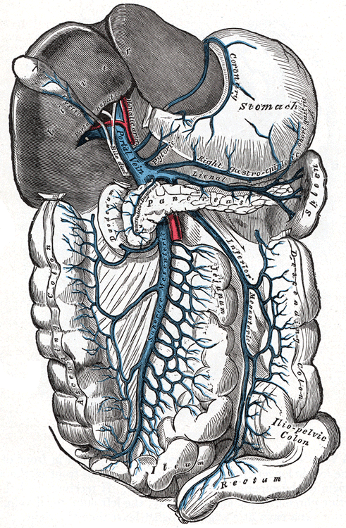
Decompensating complications
Decompensation (ascites, variceal bleed, encephalopathy, or jaundice) marks the shift to poor prognosis and should trigger transplant evaluation.
- Ascites → SBP: any new ascites needs a diagnostic paracentesis. Spontaneous bacterial peritonitis = ascitic PMN ≥ 250/mm³; most common organism E. coli. Treat empirically with a 3rd-gen cephalosporin (cefotaxime) plus IV albumin (cuts hepatorenal syndrome/mortality). It is monomicrobial — polymicrobial growth or very high ascitic protein suggests secondary (perforation). After an episode, give indefinite fluoroquinolone prophylaxis (norfloxacin/ciprofloxacin) — SBP otherwise recurs in most survivors within a year.
- Hepatic encephalopathy: ammonia-driven astrocyte swelling → asterixis. Always hunt the precipitant: GI bleed, infection, hypokalemia/alkalosis, constipation, dehydration/diuretics, TIPS, sedatives. Treat with lactulose (acidifies colon → traps NH4⁺; titrate to 2–3 soft stools/day) + rifaximin.
- Hepatorenal syndrome: functional renal failure from splanchnic vasodilation; a diagnosis of exclusion with bland sediment and no response to volume. Treat with albumin + vasoconstrictors (terlipressin, or midodrine + octreotide); definitive cure is transplant.
- HCC: cirrhosis is premalignant — screen with ultrasound ± AFP every 6 months.
Buzzwords: alcohol use / hepatitis history + hematemesis, tense ascites, or confusion with asterixis; exam shows spider angiomata, palmar erythema, splenomegaly, caput medusae.
Acute variceal bleed — the ordered answer:
- Resuscitate (2 large-bore IVs; restrictive transfusion — threshold Hgb 7, target 7–9 g/dL — improves survival; over-transfusing raises portal pressure and rebleeding).
- Octreotide (somatostatin analog → ↓ splanchnic flow / portal pressure) IV.
- Prophylactic ceftriaxone — antibiotics independently reduce mortality in any cirrhotic GI bleed.
- Urgent EGD with endoscopic band ligation (within ~12 h); TIPS if refractory.
- Non-selective beta-blocker (propranolol, nadolol, carvedilol) for primary/secondary prophylaxis — not during the acute bleed.
Other quick calls:
- New ascites → diagnostic tap with cell count, culture, and SAAG.
- Ascitic PMN ≥ 250 → cefotaxime + albumin (SBP).
- Confusion + asterixis → treat the precipitant + lactulose.
Reading the ascitic fluid: SAAG
| Feature | High SAAG ≥ 1.1 (portal HTN) | Low SAAG < 1.1 (non-portal) |
|---|---|---|
| Mechanism | Portal hypertension | Peritoneal/oncotic process |
| Classic causes | Cirrhosis, heart failure / constrictive pericarditis, Budd-Chiari | Peritoneal carcinomatosis, TB peritonitis, pancreatic, nephrotic |
| Ascitic total protein | < 2.5 cirrhosis vs > 2.5 cardiac/post-hepatic | Typically high (malignancy/TB) |
The Child-Pugh score (classes A/B/C) grades cirrhosis severity from five variables:
- P — Prothrombin time / INR
- A — Albumin
- B — Bilirubin
- A — Ascites
- E — Encephalopathy
Contrast with MELD-Na (transplant allocation priority), built from objective labs only: bilirubin, INR, creatinine, sodium. Memory hook: MELD is the lab score for the list; Child-Pugh adds the two clinical items (ascites, encephalopathy).
- Decompensation (ascites, variceal bleed, encephalopathy, jaundice) = worse survival → refer for transplant evaluation.
- MELD-Na (bilirubin, INR, creatinine, Na) prioritizes the transplant list; Child-Pugh grades severity.
- Ammonia level does NOT correlate with encephalopathy severity — diagnose and treat clinically, do not trend ammonia.
- Hepatopulmonary syndrome: liver disease + hypoxemia + intrapulmonary vasodilation → platypnea/orthodeoxia (dyspnea and desaturation worse when upright).
- Hyponatremia in cirrhosis is dilutional (ADH-driven) and is itself a poor-prognosis marker.
- NSBBs are for variceal prophylaxis — hold them in an acute bleed, and in refractory ascites or SBP with hypotension.
- Screen every cirrhotic for HCC (US ± AFP q6 months) regardless of etiology.
Practice GI now
Board-style questions, spaced-repetition flashcards, and a Socratic AI tutor — free to start.