Cellular Adaptations: Hyperplasia, Metaplasia, Dysplasia & Atrophy
A board-focused walkthrough of the four cellular adaptations — hypertrophy, hyperplasia, atrophy, and metaplasia — plus dysplasia, the preneoplastic bridge to cancer (not a true adaptation). Organized by mechanism (size vs. number vs. cell-type swap), classic organ examples, and the reversibility rule: everything is reversible until atypical cells invade through the basement membrane. Built around Step 1 vignettes (Barrett esophagus, postmenopausal bleeding) and next-best-step decisions.
What cellular adaptation means
Cellular adaptations are reversible changes in cell size, number, or phenotype that let a tissue survive a new physiologic or pathologic stress — the middle ground between the normal cell and irreversible injury/cell death. Remove the stimulus and the cell returns toward baseline; leave it excessive or persistent and some changes (especially metaplasia, and hyperplasia in select organs) can set the stage for dysplasia and ultimately neoplasia.
The four classic adaptations are hypertrophy, hyperplasia, atrophy, and metaplasia. Dysplasia is not itself an adaptation — it is disordered, preneoplastic growth — but boards group it here because metaplasia and hyperplasia can progress to it. Boards test three things: (1) telling the changes apart by mechanism (size vs. number vs. cell-type swap), (2) the classic organ examples, and (3) reversible vs. the point of no return — invasion through the basement membrane = cancer (irreversible).
- Hyperplasia = ↑ cell number (needs cells able to divide). Physiologic (breast/uterus in pregnancy, liver regeneration) or pathologic (endometrial hyperplasia, BPH).
- Hypertrophy = ↑ cell size (more structural proteins/organelles). The only option for permanent cells — cardiac & skeletal muscle, neurons. Pressure-overload LVH (HTN, aortic stenosis) re-expresses a fetal gene program (ANP/ANF, β-myosin heavy chain).
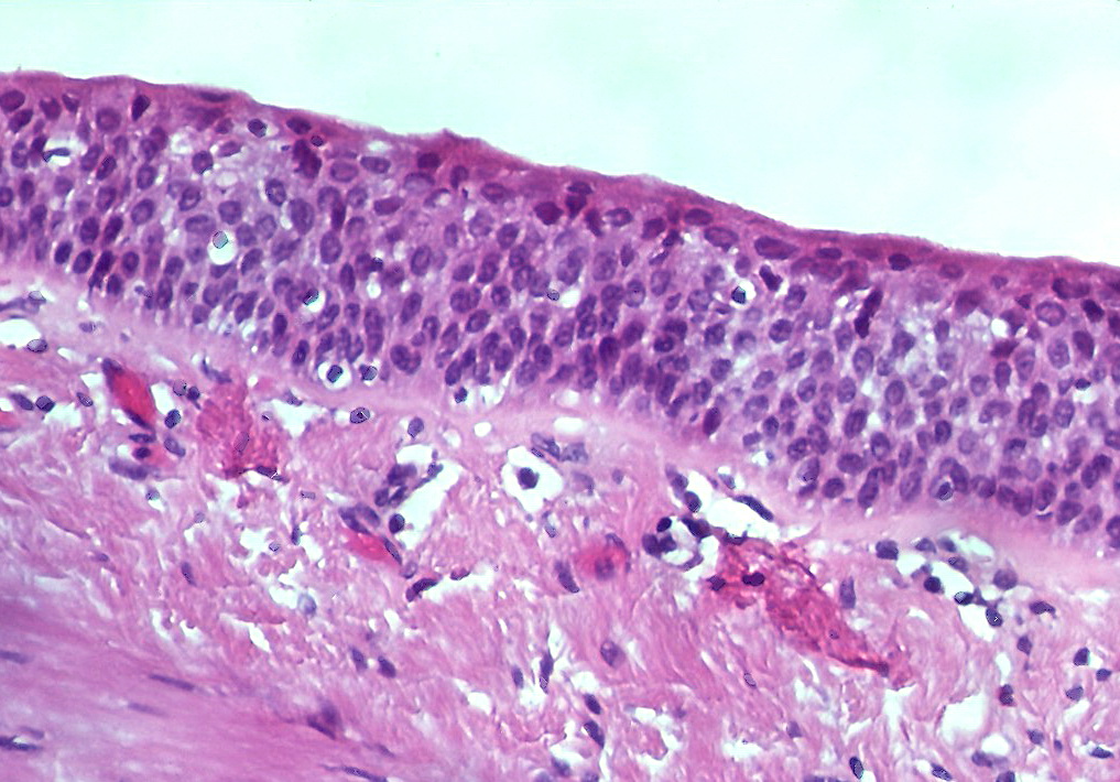
- Metaplasia = reversible swap of one mature epithelium for a hardier one, via reprogramming of stem cells (not transdifferentiation of already-mature cells).
- Atrophy = ↓ cell size/number via the ubiquitin-proteasome pathway + autophagy (disuse, denervation, ischemia, ↓ hormonal/nutritional input, pressure, aging).
- Dysplasia (not a true adaptation) = disordered, atypical growth with loss of uniformity/polarity; preneoplastic but still reversible if the insult is removed.
Side-by-side comparison
| Process | Key change | Reversible? | Classic board examples |
|---|---|---|---|
| Hyperplasia | ↑ number | Yes | Endometrial hyperplasia, BPH, Graves goiter, liver regeneration |
| Hypertrophy | ↑ size | Yes | LVH (HTN, aortic stenosis), gravid uterus, skeletal muscle in athletes |
| Metaplasia | cell-type swap | Yes | Barrett (squamous→columnar); smoker's bronchus (columnar→squamous) |
| Dysplasia | atypia/disorder | Yes (pre-invasion) | Cervical CIN, Barrett with dysplasia |
| Atrophy | ↓ size/number | Yes | Alzheimer brain, denervated/immobilized muscle, postmenopausal endometrium |
Vignette: A 55-year-old man with 10 years of reflux undergoes EGD showing salmon-colored tongues of mucosa extending proximally above the gastroesophageal junction; biopsy reveals columnar epithelium with goblet cells (intestinal metaplasia).
Dx: Barrett esophagus — GERD-driven metaplasia of the normal stratified squamous lining into intestinal-type columnar epithelium.
Why it matters / next step: It is the precursor to esophageal adenocarcinoma via the metaplasia → dysplasia → carcinoma sequence. No dysplasia → surveillance endoscopy; high-grade dysplasia → endoscopic eradication (ablation ± resection).
Buzzword: goblet cells = the defining feature of intestinal metaplasia.

Vignette: A 58-year-old obese, postmenopausal woman reports painless vaginal bleeding.
Next best step: Endometrial biopsy for tissue — postmenopausal bleeding is endometrial cancer until proven otherwise. (Transvaginal ultrasound showing a thin endometrial stripe <4 mm is an accepted alternative first test; a thickened stripe or persistent bleeding still mandates biopsy.)
Path: Unopposed estrogen (obesity, chronic anovulation/PCOS, tamoxifen, estrogen-only HRT) drives endometrial hyperplasia; hyperplasia with atypia carries the highest risk of progressing to endometrial adenocarcinoma.
Contrast trap: BPH is a hyperplasia of the prostate transition (periurethral) zone that obstructs but is not premalignant — prostate cancer arises in the peripheral zone. Not all hyperplasia is precancerous.
- Point of no return: dysplasia is reversible, but once atypical cells breach the basement membrane it is invasive carcinoma (irreversible).
- Carcinoma in situ = full-thickness dysplasia with an intact basement membrane — severe, but still not invasive.
- Metaplasia ≠ dysplasia ≠ cancer, but each step raises risk (the Barrett sequence).
- Vitamin A (retinoic acid) deficiency → squamous metaplasia of specialized epithelia (conjunctival Bitot spots, corneal keratomalacia, respiratory tract); vitamin A maintains normal epithelial differentiation.
- Hypertrophy + hyperplasia coexist where cells can divide — the gravid uterus does both.
- Atrophy is active, energy-dependent degradation (ubiquitin-proteasome + autophagy) — e.g., adrenal cortical atrophy after exogenous glucocorticoids suppress ACTH.
hyper-TROPHY → win a big trophy → cells grow in SIZE. hyper-PLASIA → PLENTY → cells grow in NUMBER.
Because permanent cells (heart, skeletal muscle, neurons) can't divide, they can only 'win a trophy': under chronic load the hypertensive/aortic-stenosis heart enlarges by adding sarcomeres and thickening its walls (hypertrophy), not by adding new myocytes (no hyperplasia).
High-yield traps to lock in
(1) Size vs. number: LVH after aortic stenosis is hypertrophy; a psoriatic plaque (acanthosis) or an estrogen-stimulated endometrium is hyperplasia.
(2) Direction of metaplasia: reflux turns esophageal squamous → columnar (Barrett), whereas smoke turns bronchial ciliated columnar → squamous — both trade a specialized epithelium for a hardier one, both lose function (e.g., mucociliary clearance), and both can progress.
(3) Reversible until invasive: removing the insult (treat reflux, stop smoking, oppose/lower estrogen) can reverse metaplasia and even dysplasia — but not invasive carcinoma.
(4) Not all hyperplasia is premalignant: BPH does not cause prostate cancer.
(5) Atrophy is active: proteasomal + autophagic degradation, not passive 'shrinkage from disuse.'
Practice Pathology now
Board-style questions, spaced-repetition flashcards, and a Socratic AI tutor — free to start.