Cell Injury, Necrosis & Apoptosis
A Step 1 walk through the cell-injury spectrum: reversible versus irreversible change, the six morphologic patterns of necrosis, and the intrinsic/extrinsic apoptosis pathways — with the buzzwords, markers, and mechanisms boards reward.
Cell injury results when a cell can no longer maintain homeostasis against a stress. The most common cause is hypoxia/ischemia, but toxins, free radicals, infections, immune reactions, and genetic or nutritional derangements all contribute. Injury lies on a spectrum: a mild or brief insult produces reversible change and the cell recovers once the stress is removed, whereas a severe or sustained insult drives the cell past a "point of no return" into irreversible injury and death. The two morphologic patterns of cell death are necrosis (always pathologic, membrane rupture, inflammation) and apoptosis (energy-dependent, programmed, non-inflammatory). Note that ischemia is more damaging than pure hypoxia: in hypoxia, blood flow persists so anaerobic glycolysis continues, whereas ischemia also cuts off substrate (glucose) delivery and lets toxic metabolites accumulate.
- Cellular (hydropic) swelling is the earliest sign of nearly all cell injury — caused by Na⁺/K⁺-ATPase failure as ATP falls.
- Other reversible changes: plasma-membrane blebbing, mitochondrial swelling, ribosomal detachment from RER (↓ protein synthesis), fatty change (steatosis), chromatin clumping.
- Two hallmarks of irreversibility: (1) severe mitochondrial damage (no ATP, even after reperfusion) and (2) membrane damage (plasma + lysosomal).
- Cytosolic Ca²⁺ influx is the key executioner — it activates phospholipases, proteases, endonucleases, and ATPases.
- Membrane rupture leaks intracellular enzymes and proteins → the clinical markers troponin (a regulatory protein, not an enzyme), CK-MB, AST/ALT, amylase, lipase, LDH.
- Free radicals (ROS) injure by lipid peroxidation, protein cross-linking, and DNA damage. Sources: reperfusion, ionizing radiation, Fenton/Haber-Weiss reactions (Fe²⁺, Cu²⁺), and drugs (CCl₄ → CCl₃· via P450, acetaminophen).
- Antioxidant defenses: superoxide dismutase, catalase, glutathione peroxidase; vitamins A, C, E; glutathione.
| Feature | Necrosis | Apoptosis |
|---|---|---|
| Trigger | Always pathologic (ischemia, toxin, trauma) | Physiologic or pathologic |
| Energy | ATP depleted | ATP required (active process) |
| Cell size | Swelling (oncosis) | Shrinkage |
| Membrane | Ruptured, leaky | Intact → blebs → apoptotic bodies |
| Inflammation | Yes (spilled contents) | No |
| DNA breakdown | Random smear | Internucleosomal "ladder" |
| Enzyme/protein leak | Yes (troponin, lipase, LDH) | No |

Necrosis is cell death with loss of membrane integrity, enzymatic digestion of the cell, and spillage of contents that triggers inflammation. Nuclear changes follow a set sequence: pyknosis (shrinkage/condensation) → karyorrhexis (fragmentation) → karyolysis (fading of basophilia from DNase digestion). Grossly and histologically, necrosis is sorted into patterns that point to a specific cause and location — pattern recognition is the high-yield boards skill. One nuance for infarcts: coagulative infarcts are pale/white in solid organs with a single end-arterial supply (heart, kidney, spleen) but red/hemorrhagic in loose or dual-supply tissues (lung, intestine, testis) and after reperfusion.
| Pattern | Classic setting | Mechanism | Buzzword / clue |
|---|---|---|---|
| Coagulative | Ischemic infarct of heart, kidney, spleen (not brain) | Protein denaturation; architecture preserved | Wedge-shaped pale infarct, "ghost cells" |
| Liquefactive | Brain infarct, bacterial abscess, pancreas | Enzymatic digestion (hydrolases/neutrophils) | Soft/cystic, pus |
| Caseous | TB, systemic fungi | Coagulative + liquefactive within a granuloma | "Cheese-like" central necrosis |
| Fat | Acute pancreatitis, breast trauma | Lipase → saponification with Ca²⁺ | Chalky-white, ↑lipase |
| Fibrinoid | Malignant HTN, vasculitis, preeclampsia | Fibrin + plasma proteins leak into damaged wall (immune complexes in vasculitis) | Bright-pink amorphous vessel wall |
| Gangrenous | Limb/GI ischemia | Dry = coagulative; Wet = + bacteria (liquefactive) | Dry vs wet gangrene |
Apoptosis is ATP-dependent, programmed cell death that removes single cells without inflammation, packaging them into apoptotic bodies that are phagocytosed (phosphatidylserine flips to the outer leaflet as the "eat-me" signal). It runs through caspase activation via two pathways. The intrinsic (mitochondrial) pathway responds to growth-factor withdrawal, DNA damage, and misfolded proteins; it is governed by the BCL-2 family — pro-apoptotic BAX/BAK versus anti-apoptotic BCL-2/BCL-XL — and ends in cytochrome c release → APAF-1 apoptosome → caspase-9. The extrinsic (death-receptor) pathway is triggered by FAS (CD95)–FASL or TNF-α, activating caspase-8. Both converge on the executioner caspases 3/6/7. Separately, cytotoxic CD8⁺ T cells kill targets via perforin/granzyme B and FAS–FASL.
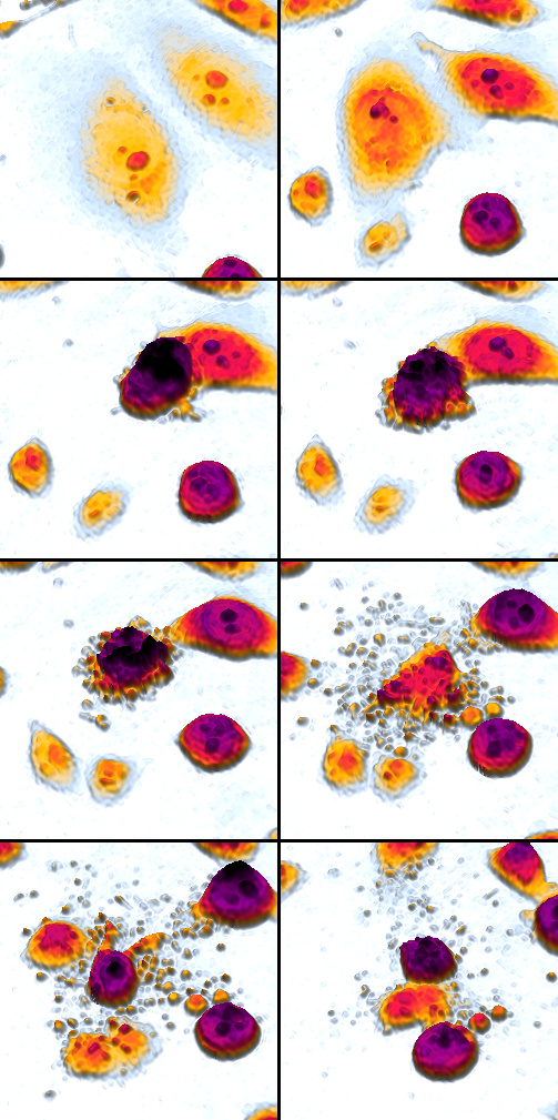
- Intrinsic → caspase-9; extrinsic → caspase-8; both converge on caspase-3 (executioner).
- BCL-2 is anti-apoptotic (keeps cytochrome c in). Overexpression via t(14;18) → follicular lymphoma (too little apoptosis → cancer).
- p53 senses DNA damage → upregulates BAX/PUMA → apoptosis. Loss of p53 (Li-Fraumeni, most cancers) → failed apoptosis.
- Too little apoptosis → cancer, autoimmunity; too much → neurodegeneration, ischemic tissue loss.
- Physiologic examples: embryogenesis (digit-webbing loss), thymic negative selection, endometrial shedding, hormone-dependent involution.
- Lab detection: annexin V (binds phosphatidylserine), TUNEL stain; gel electrophoresis shows the internucleosomal DNA ladder.
Boards test this by matching a vignette to a buzzword, then asking mechanism or next step.
- Wedge-shaped, pale, firm infarct in heart/kidney/spleen → coagulative necrosis; MI releases troponin/CK-MB; reperfusion (PCI/thrombolysis) can add free-radical reperfusion injury.
- Liquefied, cystic cavity in the brain after a stroke → liquefactive necrosis; a bacterial abscess (neutrophil hydrolases → pus) is the same pattern.
- "Cheese-like" central necrosis in a granuloma → caseous necrosis of TB or systemic fungi; hunt for acid-fast bacilli.
- Acute pancreatitis (epigastric pain to the back + ↑lipase) with chalky-white peritoneal deposits → fat necrosis. Saponification binds Ca²⁺ in the necrotic fat → dystrophic calcification (Ca²⁺ deposits in the damaged tissue with a normal serum Ca²⁺ — the classic contrast with metastatic calcification, which requires hypercalcemia); severe pancreatitis can additionally consume enough calcium to cause systemic hypocalcemia. Breast trauma gives a benign, mammographically calcified mass that mimics cancer.
- Bright-pink vessel walls in malignant hypertension, vasculitis (e.g., PAN), or preeclampsia → fibrinoid necrosis.
- Follicular lymphoma with t(14;18)/BCL-2 overexpression → decreased apoptosis.
- Apoptosis starts with "A" — ATP-dependent, Active, and Anti-inflammatory (contrast necrosis: passive, ATP-depleted, inflammatory).
- Liquefactive necrosis = brain, abscess, pancreas — soft tissues that digest themselves; coagulative is essentially everywhere else (solid-organ infarcts).
- Caspase 8 vs 9: the e8xtrinsic (death-receptor) pathway uses caspase-8; the intrinsic/mitochondrial pathway uses caspase-9.
Practice Pathology now
Board-style questions, spaced-repetition flashcards, and a Socratic AI tutor — free to start.