Acute Pancreatitis
A Step 2 CK–focused walkthrough of acute pancreatitis from trypsin-driven autodigestion through the Revised Atlanta diagnostic criteria, etiology-directed workup, and current management (early moderate goal-directed fluids, early enteral feeding, no routine antibiotics). Emphasizes next-best-step decisions for gallstone pancreatitis and infected necrosis, plus high-yield comparison tables.
Overview & Pathophysiology
Acute pancreatitis is acute inflammation of the pancreas driven by premature intra-acinar activation of trypsinogen to trypsin, which triggers a cascade of zymogen activation and autodigestion of the gland. Local injury releases inflammatory mediators that can escalate to a systemic inflammatory response (SIRS), massive third-spacing, and multi-organ failure.
Two causes account for ~70–80%: gallstones (a stone impacted at the ampulla obstructs pancreatic outflow) and chronic alcohol use. High-yield remaining causes include hypertriglyceridemia (usually TG >1000 mg/dL), hypercalcemia, post-ERCP, certain drugs (thiazides, valproate, azathioprine), trauma, and — classically in children — mumps. Always pin down the etiology: it drives definitive management (cholecystectomy for gallstones, insulin/apheresis for severe hypertriglyceridemia, stopping an offending drug).
Diagnose by the Revised Atlanta criteria — 2 of 3 required:
- Characteristic pain: acute epigastric pain radiating to the back, worse supine, relieved by leaning forward, with nausea/vomiting
- Lipase (or amylase) ≥ 3× the upper limit of normal — lipase is more sensitive/specific and stays elevated longer
- Characteristic imaging (contrast CT/MRI, or ultrasound)
Other board-tested points:
- Enzyme magnitude does NOT correlate with severity
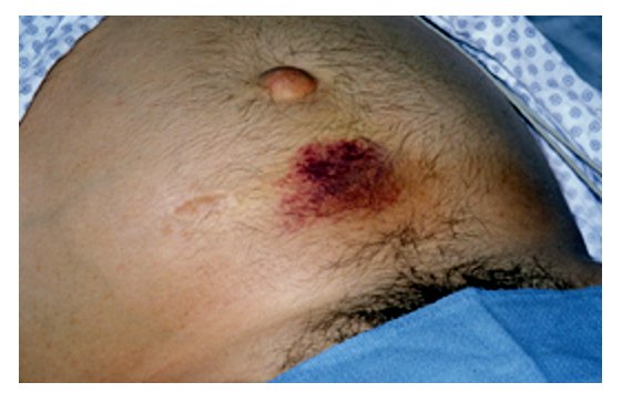
- Cullen sign (periumbilical) and Grey Turner sign (flank) ecchymoses signal retroperitoneal hemorrhage / necrotizing disease
- RUQ ultrasound in every patient to look for gallstones and biliary dilation — best initial test for etiology
- Contrast CT is not needed to make the diagnosis and underestimates necrosis early; reserve it for an unclear diagnosis or to assess complications at 48–72 hours
I GET SMASHED — classic causes of acute pancreatitis:
- I – Idiopathic
- G – Gallstones
- E – Ethanol
- T – Trauma
- S – Steroids
- M – Mumps (and other infections)
- A – Autoimmune (IgG4)
- S – Scorpion sting
- H – Hypertriglyceridemia / Hypercalcemia
- E – ERCP
- D – Drugs (thiazides, valproate, azathioprine, didanosine)
On the exam, the top two remain gallstones and alcohol — anchor to the vignette clues before reaching for a zebra.

Vignette: A 48-year-old obese woman has 6 hours of severe epigastric pain radiating to the back, nausea, and vomiting after a fatty meal. Exam: epigastric tenderness, T 38.1°C. Labs: lipase 1,200 U/L (>3× ULN), AST 180, ALT 210, total bilirubin 2.5 mg/dL, elevated alkaline phosphatase.
Diagnosis: Acute gallstone pancreatitis — an ALT >150 U/L strongly predicts a biliary origin.
Next best step:
- RUQ ultrasound to confirm stones/duct dilation; begin early goal-directed IV lactated Ringer's, analgesia, and early enteral feeding as tolerated.
- If ascending cholangitis (fever + jaundice + RUQ pain = Charcot triad) or persistent CBD obstruction → urgent ERCP with stone extraction.
- Mild gallstone pancreatitis → cholecystectomy during the same admission to prevent recurrence.
- Fluids: early, moderate goal-directed lactated Ringer's (preferred over normal saline; may lower SIRS) — resuscitate to physiologic targets (HR, MAP, urine output, falling BUN/Hct) and avoid over-resuscitation; aggressive fixed high-rate fluids increase fluid-overload/respiratory complications without benefit
- Analgesia: opioids as needed (no evidence morphine worsens outcomes)
- Nutrition: early enteral feeding within 24–72 h as tolerated; enteral > TPN (maintains gut-barrier integrity, fewer infections). Prolonged NPO/TPN is outdated
- Antibiotics: NOT routine — withhold even for sterile necrosis; give only for infected necrosis or extrapancreatic infection (e.g., cholangitis)
- Treat the cause: cholecystectomy (gallstones); insulin infusion ± apheresis, then a fibrate for long-term control (severe hypertriglyceridemia); stop the offending drug
- Severity tools: BISAP, Ranson, APACHE II; trend BUN and hematocrit to gauge resuscitation and prognosis
Vignette: A 55-year-old man hospitalized 3 weeks ago for severe necrotizing pancreatitis returns with recurrent fever, leukocytosis, and worsening abdominal pain. Contrast CT shows a walled-off peripancreatic collection containing gas bubbles.
Diagnosis: Infected walled-off necrosis — extraluminal gas implies infection by gas-forming organisms. (Image-guided FNA is rarely needed when gas is present.)
Next best step:
- Start broad-spectrum antibiotics with good pancreatic penetration — a carbapenem, or a fluoroquinolone + metronidazole.
- Delay intervention until the collection is walled off (~4 weeks), then use a minimally invasive "step-up" approach — endoscopic or percutaneous drainage first, escalating to necrosectomy only if needed, rather than upfront open surgery.
Local Complications (Revised Atlanta)
| Timing / Necrosis | Name | Key features | Management |
|---|---|---|---|
| <4 wks, no necrosis | Acute peripancreatic fluid collection | No defined wall; often resolves | Supportive |
| >4 wks, no necrosis | Pseudocyst | Encapsulated fluid, high amylase; may cause pain/mass or early satiety | Drain only if symptomatic/infected |
| <4 wks, with necrosis | Acute necrotic collection | Necrosis ± fluid, no mature wall | Supportive; antibiotics if infected |
| >4 wks, with necrosis | Walled-off necrosis | Encapsulated necrotic tissue | Step-up drainage if infected/symptomatic |

Distinguishing the Top Etiologies
| Etiology | Vignette clue | Key labs | Specific management |
|---|---|---|---|
| Gallstones | Female, fatty meal, jaundice | ALT >150, ↑alk phos; stones on US | ERCP if cholangitis; same-admission cholecystectomy |
| Alcohol | Binge or chronic use | Lipase ↑; AST:ALT >2 | Supportive; alcohol cessation, thiamine |
| Hypertriglyceridemia | Diabetes, xanthomas, lipemic serum | TG >1000; amylase may be falsely normal/low | Insulin infusion ± apheresis, then fibrate |
Practice GI now
Board-style questions, spaced-repetition flashcards, and a Socratic AI tutor — free to start.