Normal·Cardiovascular·Myocardium
Cardiac muscle — intercalated discs
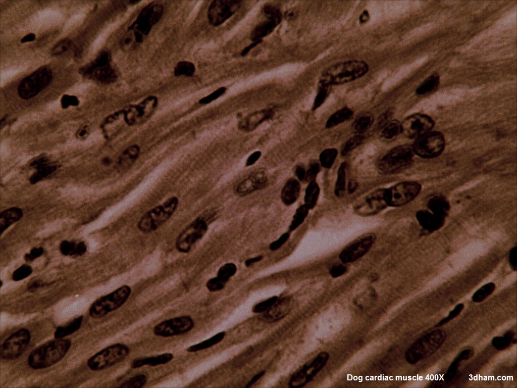
Stain: H&E·Magnification: 40x·Tissue: Cardiac muscle·3 labeled regions
Wikimedia Commons · File:Dogcardiacmuscle400.jpg · https://commons.wikimedia.org/wiki/File:Dogcardiacmuscle400.jpg (CC-licensed)
Open the interactive viewer
Sign up free to pan/zoom up to 10×, tap each labeled marker for the structure's description and clinical context, and switch to quiz mode to test yourself on every region.
Create free accountDescription
Striated branching fibers with central nuclei. Intercalated discs (gap junctions + desmosomes) couple cells electrically and mechanically — the functional syncytium of the heart.
Labeled regions (3)
- 1Intercalated disc
Stair-step junction — contains gap junctions, desmosomes, fascia adherens.
- 2Central nucleus
Single, ovoid, central — distinguishes from skeletal (peripheral).
- 3Striations
Sarcomeres confirm cardiac is striated, unlike smooth.