Pathologic·Cardiovascular·Artery (aorta/coronary)
Atherosclerosis — fibrous cap + lipid core
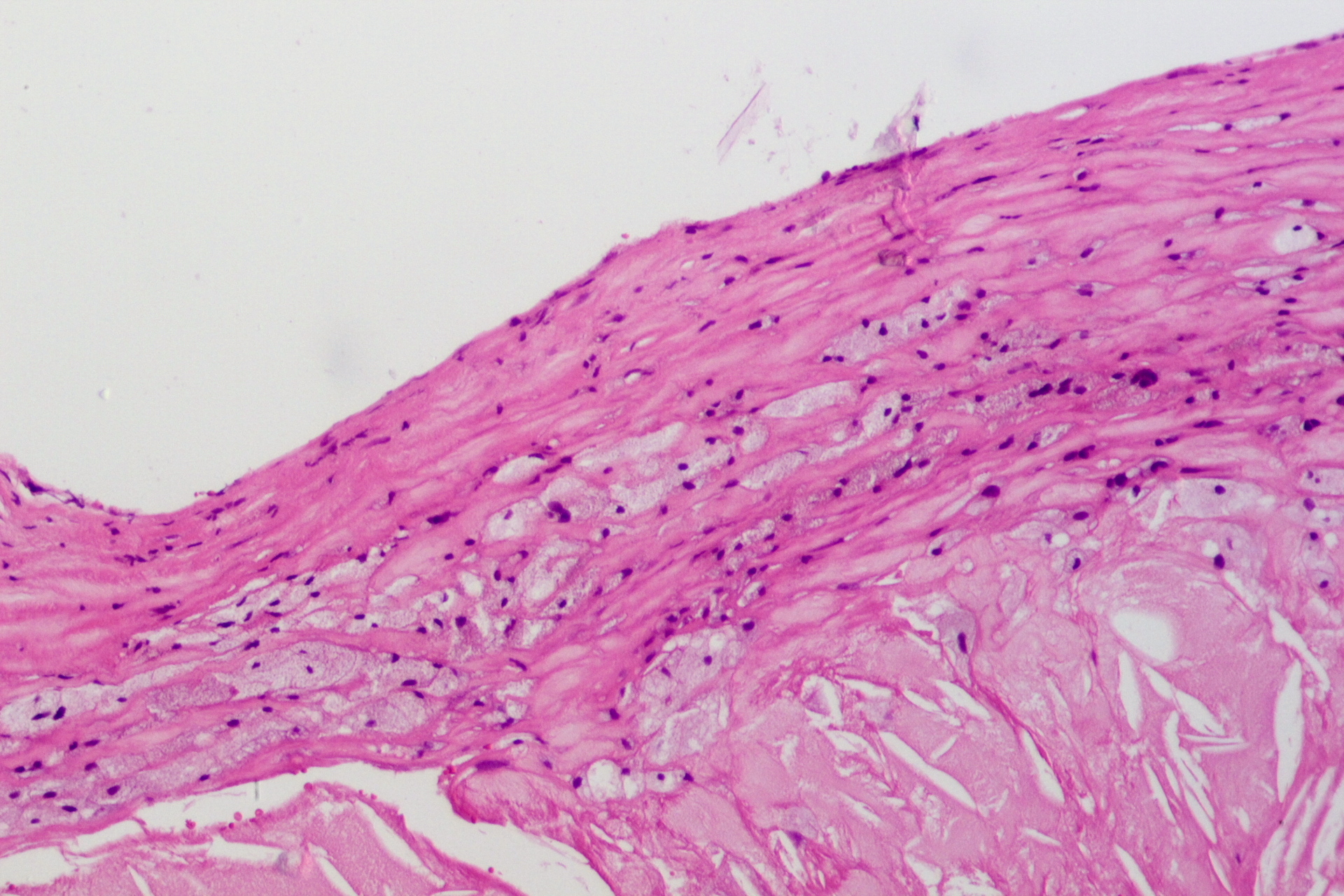
Stain: H&E·Magnification: 4x·Tissue: Intima with atheroma·4 labeled regions
Wikimedia Commons · File:Atherosclerosis,_HE_4.JPG · https://commons.wikimedia.org/wiki/File:Atherosclerosis,_HE_4.JPG (CC-licensed)
Open the interactive viewer
Sign up free to pan/zoom up to 10×, tap each labeled marker for the structure's description and clinical context, and switch to quiz mode to test yourself on every region.
Create free accountDescription
Eccentric thickening of intima: fibrous cap (smooth muscle + collagen) covering a necrotic lipid core (cholesterol clefts, foamy macrophages). Cap rupture exposes thrombogenic core → MI / stroke.
Labeled regions (4)
- 1Fibrous cap
Smooth muscle cells + collagen overlying the core. Thin caps + inflammation = rupture-prone (vulnerable plaque).
- 2Necrotic lipid core
Cholesterol clefts, foam cells (lipid-laden macrophages), debris. Highly thrombogenic if exposed.
- 3Tunica media
Smooth muscle wall — thinned + remodeled overlying plaques. Internal/external elastic laminae often disrupted.
- 4Lumen (narrowed)
Critical stenosis (>70%) drives stable angina / claudication. Acute thrombosis on rupture drives MI.