Vertigo & Vestibular Disorders
A Step 2 CK high-yield lesson on vertigo that anchors on the peripheral-vs-central distinction, then walks through BPPV, vestibular neuritis/labyrinthitis, Ménière disease, and posterior-circulation stroke via buzzword vignettes and next-best-step decisions. Includes a comparison table plus the INFARCT (HINTS) and COWS mnemonics.
Overview: The Peripheral vs Central Split
Vertigo is the false sensation of movement (usually spinning) from asymmetry in the vestibular system — not lightheadedness or presyncope. On the boards, the task is almost always to separate peripheral vertigo (inner ear: labyrinth, semicircular canals, CN VIII) from central vertigo (brainstem/cerebellum), because a benign inner-ear problem and a posterior-circulation stroke can look identical.
Peripheral causes are more common, usually benign, but intensely symptomatic. Central causes are dangerous and demand imaging. Peripheral clues: vertigo that is brief and positional (BPPV) or continuous for days (neuritis); horizontal, unidirectional nystagmus that suppresses with visual fixation; often hearing symptoms; and — in the continuous presentation — an abnormal head-impulse test. Central red flags: milder but sustained imbalance, direction-changing, vertical, or pure-torsional nystagmus, inability to stand/walk, and any focal neuro deficit (dysarthria, dysphagia, diplopia).
The four disorders tested repeatedly: BPPV, vestibular neuritis/labyrinthitis, Ménière disease, and central (stroke).
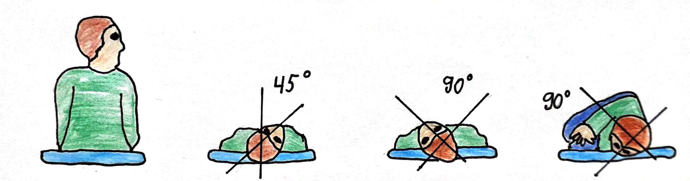
- BPPV — brief (<1 min) vertigo triggered by head-position change (rolling in bed, looking up); Dix-Hallpike provokes upbeat-torsional nystagmus with latency and fatigability; treat with the Epley maneuver. No hearing loss.
- Vestibular neuritis — acute, continuous vertigo lasting days, often post-viral; horizontal unidirectional nystagmus, no hearing loss. Add hearing loss → labyrinthitis.
- Ménière disease — recurrent spells (20 min–hours) of vertigo + fluctuating low-frequency SNHL + tinnitus + aural fullness; endolymphatic hydrops.
- Central (stroke) — sudden continuous vertigo + vascular risk factors; direction-changing/vertical nystagmus, severe gait ataxia, or focal signs (Wallenberg: Horner, dysphagia, crossed sensory loss).
- HINTS exam applies only to continuous vertigo (acute vestibular syndrome) with nystagmus, and only when performed by a trained examiner — done correctly it outperforms early MRI-DWI for posterior stroke.

Comparison Table
| Feature | BPPV | Vestibular neuritis | Ménière | Central (stroke) |
|---|---|---|---|---|
| Duration | Seconds (<1 min), episodic | Days, continuous | 20 min–hours, recurrent | Continuous, persistent |
| Trigger | Head-position change | Spontaneous (post-viral) | Spontaneous | Spontaneous |
| Hearing loss | No | No (yes = labyrinthitis) | Yes, fluctuating low-freq SNHL | Usually no (AICA can) |
| Nystagmus | Upbeat-torsional, fatigable | Horizontal, unidirectional | Horizontal | Direction-changing / vertical |
| Fixation suppresses? | n/a (positional) | Yes | Yes | No |
| Key test | Dix-Hallpike | Head-impulse (abnormal) | Audiometry (low-freq SNHL) | HINTS central; MRI-DWI |
| Treatment | Epley maneuver | Supportive + vestibular rehab | Low-salt diet + thiazide | Acute stroke pathway |
Vignette: A 58-year-old woman has recurrent ~20-second spinning episodes whenever she rolls over in bed or tilts her head back. No hearing loss or tinnitus; neuro exam normal. Lowering her into the Dix-Hallpike position reproduces vertigo with upbeating-torsional nystagmus after a brief latency, fatiguing on repetition.
Diagnosis: Benign paroxysmal positional vertigo (posterior semicircular canal canalithiasis).
Next best step: Perform the Epley (canalith-repositioning) maneuver — first-line and often curative. Vestibular suppressants (meclizine) are not the answer and can impair central compensation; reserve brief use for severe acute symptoms only. Imaging is unnecessary with a classic positional history and a normal neuro exam.
Vignette: A 70-year-old man with hypertension and diabetes has sudden, continuous vertigo, vomiting, and unsteadiness for 3 hours. Nystagmus changes direction with gaze, and he cannot stand without falling. Head-impulse test is normal, and there is vertical skew on cover testing.
Diagnosis: Central vertigo — posterior-circulation (cerebellar/brainstem) stroke until proven otherwise. All three HINTS components are central (INFARCT).
Next best step: Activate the stroke workup and obtain MRI with diffusion — but note DWI is falsely negative in up to ~20% of posterior strokes in the first 24–48 h, so a reassuring early scan does not exclude stroke and exam/HINTS drives decisions. A normal head-impulse test during acute continuous vertigo is a danger sign, not reassurance.
HINTS → 'INFARCT' — findings that point central in acute continuous vertigo:
- Impulse Normal — normal head-impulse test
- Fast-phase Alternating — direction-changing nystagmus
- Refixation on Cover Test — vertical skew deviation
Any one central HINTS finding = worry about stroke. (A peripheral lesion instead gives an abnormal head-impulse test — a visible corrective/catch-up saccade toward the lesioned side.)
Caloric testing → 'COWS': Cold Opposite, Warm Same — names the direction of the fast phase of nystagmus (cold water → fast phase beats to the opposite side; warm → same side).
- BPPV: Epley maneuver; recurrences are common; Brandt-Daroff home exercises for refractory cases.
- Vestibular neuritis / labyrinthitis: supportive — short-course vestibular suppressants (meclizine, benzodiazepines) + antiemetics for ≤3 days, then early vestibular rehabilitation; corticosteroids are sometimes given, but the evidence for benefit is mixed.
- Ménière: low-sodium diet, limit caffeine/alcohol; thiazide diuretic; treat acute attacks with meclizine/antiemetics; refractory → intratympanic steroids, then intratympanic gentamicin (ablative).
- Central: admit, stroke team, imaging, treat the vascular cause.
- Pearl: Prolonged vestibular suppressants delay central compensation — use sparingly and stop early.
Vignette: A 45-year-old woman reports several months of recurrent 1–2 hour vertigo spells with roaring tinnitus, a sense of ear fullness, and hearing that worsens during attacks. Audiometry shows low-frequency sensorineural hearing loss in one ear.
Diagnosis: Ménière disease (endolymphatic hydrops).
Next best step: Begin sodium restriction and a thiazide diuretic, and counsel to limit caffeine/alcohol; treat acute vertigo with meclizine/antiemetics. Refer to ENT/audiology; escalate to intratympanic therapy only if symptoms remain refractory.
Practice ENT (Otolaryngology) now
Board-style questions, spaced-repetition flashcards, and a Socratic AI tutor — free to start.