Uveitis & Scleritis
A Step 2 CK walkthrough of uveitis and scleritis: recognizing each painful red eye, tying it to its systemic disease (HLA-B27, sarcoid, RA, GPA), using the phenylephrine test to separate scleritis from episcleritis, and choosing the correct next-best-step management.
Overview: two red eyes the boards love
Uveitis is inflammation of the uveal tract (iris, ciliary body, choroid), classified by site: anterior (iritis/iridocyclitis — most common), intermediate, posterior (chorioretinitis), or panuveitis. Scleritis is inflammation of the sclera itself, with congestion of the deep episcleral vascular plexus (episcleritis involves only the superficial plexus). Both present as a painful red eye, and Step 2 CK contrasts them against benign mimics (conjunctivitis, episcleritis).
The exam rewards two moves: (1) link the eye to a systemic disease — HLA-B27 spondyloarthropathies and sarcoidosis for anterior uveitis; rheumatoid arthritis (RA) and granulomatosis with polyangiitis (GPA) for scleritis; and (2) pick the next best step, which is prompt ophthalmology referral plus the correct anti-inflammatory ladder. Missing scleritis risks scleral necrosis and globe perforation; missing uveitis risks posterior synechiae, cataract, and secondary glaucoma.
- Anterior uveitis = painful red eye, photophobia, ciliary (circumcorneal) flush; slit lamp shows cells & flare + keratic precipitates; severe cases form a hypopyon
- Consensual photophobia: shining light in the unaffected eye triggers pain in the affected eye (via the consensual light reflex) — classic for iritis
- Pupil is small/constricted or irregular (ciliary spasm); posterior synechiae (iris adherent to lens) are a key complication
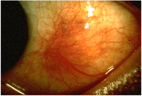
- Scleritis = severe, deep, boring pain radiating to brow/jaw, wakes patient from sleep, worse with eye movement; bluish-violaceous sclera
- Phenylephrine test — scleral vessels do NOT blanch (deep); episcleritis DOES blanch (superficial): the single best discriminator
- Episcleritis = mild, sectoral redness, little/no pain, normal vision, self-limited
- Necrotizing scleritis (incl. scleromalacia perforans, the non-inflamed form seen in longstanding RA) is a systemic/surgical emergency
Anterior uveitis vs. scleritis vs. episcleritis
| Feature | Anterior uveitis | Scleritis | Episcleritis |
|---|---|---|---|
| Pain | Aching + photophobia | Severe, boring, wakes from sleep | Mild or none |
| Redness | Ciliary flush | Diffuse, deep, violaceous | Sectoral, superficial |
| Vision | Decreased | Often decreased | Normal |
| Phenylephrine | — | Does NOT blanch | Blanches |
| Slit lamp | Cells/flare, KPs, hypopyon | Scleral edema | Normal |
| Top associations | HLA-B27, sarcoid, JIA | RA, GPA | Idiopathic |
| Treatment | Topical steroid + cycloplegic | Systemic NSAID/steroid | Lubricants, reassurance |
Vignette: A 26-year-old man with months of low back stiffness that improves with exercise and worsens with rest presents with a painful, red, light-sensitive right eye and blurred vision. Exam shows circumcorneal (ciliary) flush; slit lamp reveals anterior-chamber cells and flare with keratic precipitates.
Diagnosis: Acute anterior uveitis associated with HLA-B27 ankylosing spondylitis.
Next best step: Urgent ophthalmology referral; start topical corticosteroids plus a cycloplegic/mydriatic (e.g., cyclopentolate) to control inflammation, relieve ciliary spasm, and prevent posterior synechiae. Work up the systemic cause (sacroiliac imaging; HLA-B27 supports but is not required for the ophthalmic diagnosis).

"PAIR" = the HLA-B27 spondyloarthropathies that cause recurrent acute anterior uveitis:
- Psoriatic arthritis
- Ankylosing spondylitis
- IBD-associated (enteropathic) arthritis
- Reactive arthritis
Also high-yield for anterior uveitis: sarcoidosis, and in a child with pauciarticular, ANA-positive juvenile idiopathic arthritis (JIA). JIA-associated uveitis is often chronic and painless (white eye), so these children need routine slit-lamp screening to avoid silent vision loss.
Vignette: A 58-year-old woman with long-standing rheumatoid arthritis reports 4 days of severe, deep, boring left-eye pain radiating to her jaw that wakes her at night and worsens with eye movement. The sclera has a violaceous hue and does not blanch after topical phenylephrine.
Diagnosis: Scleritis (RA-associated; consider GPA if sinus/nasal or renal disease is present).
Next best step: Urgent ophthalmology referral plus systemic therapy — oral NSAIDs for mild non-necrotizing disease, escalating to systemic corticosteroids ± immunosuppression (e.g., for necrotizing or GPA-related disease). Topical steroids alone are inadequate. Screen with RF/anti-CCP and ANCA, and treat the underlying autoimmune disease.
- Conjunctivitis: diffuse redness, discharge/itch, NO true pain, photophobia, or vision loss — not uveitis/scleritis
- Acute angle-closure glaucoma: fixed mid-dilated pupil, cloudy cornea, nausea/vomiting, high IOP — vs. the small/constricted pupil of anterior uveitis
- Episcleritis blanches with phenylephrine; scleritis does not — the most testable single fact
- New scleritis → screen for RA and GPA (RF/anti-CCP, ANCA)
- Posterior uveitis (floaters, painless vision loss): think toxoplasmosis, CMV retinitis in AIDS (CD4 <50), syphilis, sarcoidosis
- Never give topical steroids for a red eye before excluding HSV keratitis (dendritic ulcer on fluorescein) — steroids worsen it
Practice Ophthalmology now
Board-style questions, spaced-repetition flashcards, and a Socratic AI tutor — free to start.