Transplant Immunology & Rejection
A Step 1 high-yield lesson on transplant immunology covering graft types, HLA/MHC-based allorecognition (direct vs indirect), the four rejection patterns (hyperacute, acute cellular, acute humoral, chronic) plus GVHD, and the molecular targets of immunosuppression. Emphasizes the board-tested defect-to-disease correlations (preformed antibodies, C4d, cardiac allograft vasculopathy, bronchiolitis obliterans). Verified accurate; highYield now carries the core immunosuppressant mechanisms, and the three-signal framing was tightened so calcineurin inhibitors correctly map to IL-2 production rather than the IL-2 response.
The Immunologic Basis of Rejection
Grafts are classified by donor–recipient relationship: an autograft is self-to-self, an isograft (syngeneic) is between identical twins, an allograft is between genetically different members of the same species (the usual clinical transplant), and a xenograft is between species. Rejection is fundamentally the recipient immune system recognizing donor MHC (HLA) molecules as non-self.
- MHC class I = HLA-A, B, C — on all nucleated cells, presents to CD8+ T cells.
- MHC class II = HLA-DR, DP, DQ — on antigen-presenting cells, presents to CD4+ T cells.
- HLA-DR matching most strongly predicts graft survival (importance roughly DR > B > A); ABO compatibility is mandatory.
Two pathways of allorecognition drive different rejection timelines:
- Direct — recipient T cells recognize intact donor MHC displayed on donor 'passenger' APCs. The precursor frequency of alloreactive T cells is unusually high (~1–10%), which is why this pathway powers acute rejection.
- Indirect — recipient APCs engulf shed donor antigens and present donor-derived peptides on recipient (self) MHC to recipient CD4+ T cells. This slow, ongoing process underlies chronic rejection.

- Rejection targets donor HLA; HLA-DR match matters most for survival; ABO compatibility is required.
- Hyperacute (minutes–hours): preformed recipient antibodies (anti-ABO or anti-HLA) → type II hypersensitivity → complement + vascular thrombosis. Prevented by ABO matching and crossmatch.
- Acute (weeks–months): two flavors — cellular (CD8/CD4 T cells vs donor MHC; interstitial lymphocytic infiltrate, tubulitis, endarteritis) and humoral/antibody-mediated (C4d deposition in peritubular capillaries). Both are typically reversible with immunosuppression.
- Chronic (months–years): dominated by indirect allorecognition; intimal smooth-muscle proliferation, vascular fibrosis, parenchymal atrophy. Irreversible.
- GVHD: donor T cells attack an immunocompromised host; type IV; hits skin, gut, liver; classic after allogeneic bone-marrow/HSC transplant.
- Memory hook: Direct pathway → Acute; Indirect pathway → Chronic.
- Crossmatch = recipient serum + donor lymphocytes → detects preformed anti-HLA antibodies before surgery.
- Immunosuppression core: cyclosporine→cyclophilin and tacrolimus (FK506)→FKBP both inhibit calcineurin → block NFAT → ↓ IL-2 production; sirolimus→FKBP→mTOR (blocks IL-2 signaling); basiliximab = anti-CD25 (IL-2R); belatacept (CTLA-4-Ig) blocks B7 (CD80/86) costimulation.
The Four Rejection Patterns (Compare & Contrast)
| Type | Onset | Mechanism | Key findings / histology | Reversible? |
|---|---|---|---|---|
| Hyperacute | Minutes–hours | Preformed anti-donor Ab (ABO/HLA) + complement (type II) | Graft mottled/cyanotic; fibrinoid necrosis and thrombosis of vessels | No — graft must be removed |
| Acute cellular | Weeks–months | Recipient CD8/CD4 T cells vs donor MHC (direct) | Dense interstitial mononuclear infiltrate, tubulitis, endarteritis | Yes |
| Acute humoral | Weeks–months | De novo anti-donor antibodies | C4d+ in peritubular capillaries, capillaritis, neutrophils | Yes (harder) |
| Chronic | Months–years | Indirect allorecognition; Ab + cell-mediated | Intimal thickening/fibrosis, organ-specific atrophy | No |
| GVHD | Variable | Donor T cells vs host (type IV) | Skin rash, cholestatic LFTs, diarrhea; epithelial apoptosis | — (prevent/immunosuppress) |
Case: A woman with end-stage renal disease and a history of multiple pregnancies and prior transfusions (high panel-reactive antibody) receives a deceased-donor kidney. Within minutes of reperfusion, the graft becomes mottled, cyanotic, and flaccid, and produces no urine. Biopsy shows thrombosed vessels with fibrinoid necrosis and neutrophils.
Defect → disease: Preformed recipient antibodies against donor ABO or HLA antigens bind graft endothelium → complement activation (type II hypersensitivity) → widespread microvascular thrombosis. This is hyperacute rejection.
- Board triggers: prior pregnancies, transfusions, or transplants (sensitization); failure on the table or within hours.
- Management: none salvages it — the graft is explanted. It is prevented by ABO matching and a pre-transplant crossmatch.
Case: Three weeks after an allogeneic hematopoietic stem-cell transplant for AML, a patient develops a maculopapular rash on the palms and soles, watery diarrhea, and a rising bilirubin with cholestatic LFTs.
Defect → disease: Immunocompetent donor T cells recognize host tissue as foreign — graft-versus-host disease (GVHD), a type IV reaction. Billingham's requirements: (1) graft contains immunocompetent cells, (2) host is immunocompromised/unable to reject, (3) host expresses antigens foreign to the graft (MHC mismatch).
- Target triad: skin, GI tract, liver.
- A related pitfall: transfusion-associated GVHD from non-irradiated blood products in immunocompromised patients — prevented by irradiating blood.
- Silver lining: the same donor T cells produce a beneficial graft-versus-leukemia effect.
A real, board-tested naming trick for the T-cell immunosuppressants:
- Cyclosporine binds cyclophilin (both start with cyclo-).
- Tacrolimus (FK506) binds FKBP (FK-Binding Protein — literally named for it).
- Both drug–protein complexes inhibit calcineurin → block NFAT → ↓ IL-2 transcription → less T-cell proliferation.
- Sirolimus also binds FKBP, but instead inhibits mTOR — it blocks IL-2 signal transduction / cell-cycle progression, not IL-2 production.
So: same partner protein (FKBP) for tacrolimus and sirolimus, different downstream target (calcineurin vs mTOR).
Cells & Molecules → Function → Transplant Relevance
| Cell / molecule | Core function | Why it matters in transplant |
|---|---|---|
| MHC I (HLA-A/B/C) | Presents endogenous peptide to CD8+ T cells | The self/non-self signal read by CTLs in acute cellular rejection |
| MHC II (HLA-DR/DP/DQ) | Presents to CD4+ T cells on APCs | HLA-DR match most predicts graft survival; ignites the alloresponse |
| CD8+ cytotoxic T cell | Kills via perforin/granzyme and Fas | Direct destruction of donor graft cells |
| CD4+ helper T cell | Secretes IL-2 and cytokines; helps B cells | Orchestrates both cellular and antibody-mediated rejection |
| Preformed IgG (anti-ABO/HLA) | Fixes complement (type II) | Cause of hyperacute rejection |
| C4d | Complement split product on endothelium | Tissue marker of antibody-mediated (humoral) rejection |
| B7 (CD80/86) – CD28 | Costimulation signal 2 | Blocked by belatacept (CTLA-4-Ig) → T-cell anergy |
| IL-2 / IL-2R (CD25) | Drives T-cell clonal expansion | Calcineurin inhibitors ↓ IL-2 production; sirolimus blocks IL-2R→mTOR signaling; basiliximab = anti-CD25 (blocks the receptor) |
Blocking Rejection: The Three-Signal Framework
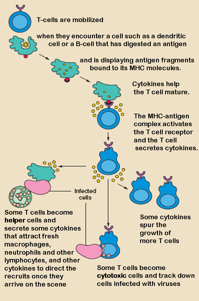
T-cell activation — and therefore rejection — depends on three signals, each an immunosuppressant target:
- Signal 1 — TCR recognizes peptide–MHC. Blocked by muromonab/anti-CD3 and lymphocyte-depleting agents (anti-thymocyte globulin, alemtuzumab).
- Signal 2 — costimulation via B7 (CD80/86) → CD28. Without it, the T cell becomes anergic. Belatacept (CTLA-4-Ig) competitively blocks B7.
- Signal 3 — IL-2-driven clonal expansion. Calcineurin inhibitors (cyclosporine, tacrolimus) act on the signal-1/2 transduction pathway to block IL-2 production (via NFAT); basiliximab blocks the IL-2 receptor (CD25); mTOR inhibitors (sirolimus) block IL-2 downstream signaling / cell-cycle progression.
Other anti-proliferatives: mycophenolate inhibits IMP dehydrogenase (blocks de novo guanine synthesis, selectively starving lymphocytes), azathioprine is a purine antimetabolite (via 6-mercaptopurine), and glucocorticoids broadly suppress NF-κB. Clinically, these regimens are excellent at preventing and reversing acute (T-cell) rejection but largely fail against chronic rejection, whose fibrotic vascular remodeling is refractory to immunosuppression.
Practice Immunology now
Board-style questions, spaced-repetition flashcards, and a Socratic AI tutor — free to start.