Thyroid & Adrenal Physiology
A high-yield Step 1 walkthrough of the HPT and HPA axes — thyroid hormone synthesis and its drug/enzyme targets, peripheral T4→T3 activation and TBG effects, adrenal zonation and cortisol/aldosterone actions, the three CAH enzyme deficiencies, and the feedback logic behind Graves, Hashimoto, Addison, Cushing, and Conn. Built around the three tested lenses: synthesis step, feedback loop, and peripheral action.
Two axes, one logic
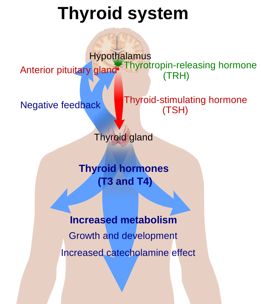
Two endocrine axes dominate Step 1: the hypothalamic–pituitary–thyroid (HPT) axis and the hypothalamic–pituitary–adrenal (HPA) axis. Both share the same architecture — a hypothalamic releasing hormone drives an anterior-pituitary trophic hormone, which drives a peripheral gland, whose product feeds back negatively on both upstream levels.
- HPT: TRH → TSH → T4/T3; free T3/T4 inhibit TSH and TRH.
- HPA: CRH → ACTH → cortisol; cortisol inhibits ACTH and CRH.
Board questions attack one of three points: (1) a synthesis/enzyme step and the drug or defect that blocks it; (2) the feedback loop (predict the trophic hormone when the gland fails or a tumor secretes autonomously — trophic hormone always moves opposite to the end-hormone in primary disease); or (3) a peripheral action (metabolism, β-adrenergic permissiveness, salt handling). Treat these as three lenses and most items become deduction, not memorization.
One wiring detail that generates questions: TRH also stimulates prolactin, so primary hypothyroidism (high TRH) can cause hyperprolactinemia and galactorrhea.
Thyroid synthesis — the assembly line (and where drugs cut it)
- Trapping: the Na⁺/I⁻ symporter (NIS) on the basolateral membrane pumps iodide into the follicular cell against its gradient — secondary active transport powered by the Na⁺/K⁺-ATPase. Blocked by perchlorate, pertechnetate, thiocyanate.
- Efflux to colloid across the apical membrane via pendrin.
- Organification + coupling by thyroid peroxidase (TPO): oxidizes iodide, iodinates tyrosines on thyroglobulin (→ MIT, DIT), then couples them — DIT + DIT → T4, MIT + DIT → T3. Blocked by thionamides (methimazole, PTU).
- Endocytosis + proteolysis: colloid is resorbed; lysosomal proteases release T4 and T3. The gland secretes mostly T4 (~90%).
- Peripheral activation: 5′-deiodinase converts the T4 prohormone to the more potent T3 in target tissues. PTU additionally blocks this peripheral 5′-deiodinase — methimazole does not.
Wolff–Chaikoff: a large iodide load transiently shuts down organification (protective autoregulation). Jod–Basedow: the mirror image — iodine given to a nodular/autonomous gland triggers hyperthyroidism.
- T4 is the reservoir/prohormone; T3 is the biologically active hormone (several-fold more potent). ~90% of thyroid secretion is T4; most circulating T3 comes from peripheral 5′-deiodination of T4.
- Reverse T3 (rT3) is inactive; illness/fasting shunts T4 → rT3 ("euthyroid sick syndrome": ↓T3, ↑rT3).
- Deiodinases are selenium-dependent (selenocysteine).
- Most hormone is protein-bound (TBG ≫ transthyretin, albumin); only free hormone is active.
- The TBG trap: ↑TBG (pregnancy, OCPs, estrogen) raises total T4 but free T4 and TSH stay normal → euthyroid. ↓TBG (nephrotic syndrome, liver failure) does the reverse.
- Mechanism: T3 binds a nuclear receptor on a thyroid response element → ↑ gene transcription.
- Actions: ↑ basal metabolic rate via ↑ Na⁺/K⁺-ATPase (↑O₂ consumption, thermogenesis); ↑ β1-adrenergic receptor expression (permissive → tachycardia, tremor); ↑ glycogenolysis, gluconeogenesis, lipolysis; required for CNS maturation and bone growth in infants.
- Screening: because free T3/T4 suppress TSH, in primary thyroid disease TSH moves opposite to the hormone — TSH is the best screening test.
- Graves disease (hyperthyroid): a stimulating IgG (thyroid-stimulating immunoglobulin, TSI) binds and activates the TSH receptor → diffuse goiter, ↑T4/T3 with suppressed TSH. The same antibody stimulates retro-orbital and dermal fibroblasts to deposit glycosaminoglycans, producing exophthalmos and pretibial myxedema — findings unique to Graves precisely because they are antibody-driven, not hormone-driven. Vignette: young woman, heat intolerance, weight loss, palpitations, proptosis.
- Hashimoto thyroiditis (hypothyroid): anti-TPO / antithyroglobulin antibodies + lymphocytic infiltration (Hürthle cells, germinal centers) destroy the gland → ↑TSH, ↓T4. A brief hashitoxicosis hyperthyroid phase can occur as stored hormone leaks. Associated with HLA-DR3/DR5 and increased risk of thyroid (MALT) lymphoma.
- Congenital hypothyroidism (cretinism): iodine deficiency or dysgenesis → the classic 6 P's — Pot-bellied, Pale, Puffy-faced, Protruding umbilicus, Protuberant tongue, Poor brain (intellectual disability) — underscoring thyroid hormone's role in CNS and skeletal maturation.
Adrenal cortex — three zones, three products, one medulla
The cortex is layered G-F-R (Glomerulosa, Fasciculata, Reticularis), making Salt, Sugar, Sex; the medulla (neural-crest chromaffin cells) makes catecholamines.
- Glomerulosa → aldosterone, driven by angiotensin II and K⁺ (not ACTH).
- Fasciculata → cortisol, driven by ACTH.
- Reticularis → androgens (DHEA), driven by ACTH.
All steroids begin from cholesterol; the rate-limiting step is desmolase (cholesterol side-chain cleavage, StAR-dependent), stimulated by ACTH.
Cortisol does a lot ("A BIG FIB"): ↑Appetite; maintains Blood pressure by upregulating α1 receptors (permissive for catecholamine vasoconstriction); ↑Insulin resistance (diabetogenic); ↑Gluconeogenesis, lipolysis, proteolysis; inhibits Fibroblasts (thin skin, striae, poor healing); Inflammatory/immune suppression (blocks phospholipase A2 and NF-κB; ↓ leukotrienes/prostaglandins/histamine; neutrophilia from demargination but ↓ eosinophils/lymphocytes); ↓ Bone formation. Cortisol follows a diurnal rhythm, peaking in the early morning.
- Aldosterone acts on principal cells of the cortical collecting duct: inserts ENaC and Na⁺/K⁺-ATPase → Na⁺ (and water) reabsorption and K⁺ secretion; it also drives H⁺ secretion by α-intercalated cells. Excess → hypokalemia + metabolic alkalosis.
- Triggers for aldosterone: angiotensin II and hyperkalemia (K⁺ directly depolarizes glomerulosa cells); ACTH is only a minor/permissive stimulus.
- RAAS chain: ↓renal perfusion → renin (JG cells) → angiotensin I → ACE (lung) → angiotensin II → vasoconstriction, aldosterone, ADH, thirst, and efferent-arteriole constriction that preserves GFR (why ACE inhibitors can drop GFR).
- ACTH is cleaved from POMC, which also yields MSH — chronically high ACTH (primary adrenal failure, ectopic ACTH) causes hyperpigmentation.
- Cortisol feedback acts at both pituitary and hypothalamus; exogenous glucocorticoids suppress CRH/ACTH → adrenal atrophy → never stop abruptly.
- Congenital adrenal hyperplasia (CAH): any cortisol-blocking enzyme defect → ↓cortisol → loss of feedback → ↑ACTH → adrenal hyperplasia with precursor shunting.
CAH enzyme deficiencies — the money table
| Feature | 21-hydroxylase (~90%, most common) | 11β-hydroxylase | 17α-hydroxylase |
|---|---|---|---|
| Cortisol | ↓ | ↓ | ↓ |
| Mineralocorticoid effect | ↓ aldosterone → salt wasting | ↑ (11-deoxycorticosterone, DOC) | ↑ (DOC) |
| Blood pressure / K⁺ | ↓ BP, ↑ K⁺ (hypotension, hyperkalemia) | ↑ BP, ↓ K⁺ | ↑ BP, ↓ K⁺ |
| Androgens | ↑ — virilization, ambiguous genitalia in girls | ↑ — virilization | ↓ — no puberty; ambiguous in boys / phenotypic female |
| Key lab marker | ↑ 17-hydroxyprogesterone | ↑ 11-deoxycorticosterone & 11-deoxycortisol | ↓ androgens & cortisol, ↑ mineralocorticoids |

- Primary adrenal insufficiency (Addison): autoimmune (or TB) destruction of the cortex → both cortisol AND aldosterone fall. Aldosterone loss → hyponatremia, hyperkalemia, hypotension, metabolic acidosis; loss of cortisol feedback → ↑ACTH → hyperpigmentation (POMC/MSH).
- Secondary insufficiency (↓ACTH — pituitary failure or abrupt steroid withdrawal): cortisol low but aldosterone preserved (RAAS is intact) → no hyperkalemia and no hyperpigmentation (ACTH is low). This mineralocorticoid-and-pigment split is the classic tested discriminator.
- Cushing syndrome (cortisol excess): central obesity, moon facies, striae, proximal weakness, hyperglycemia, osteoporosis. Localize with ACTH + dexamethasone suppression — exogenous steroids (↓ACTH; most common overall) vs Cushing disease (pituitary adenoma: ACTH high, suppresses with high-dose dex) vs ectopic ACTH (e.g., small cell lung: high ACTH, no suppression) vs adrenal tumor (↓ACTH).
- Conn syndrome (primary hyperaldosteronism): autonomous aldosterone → hypertension, hypokalemia, metabolic alkalosis, and LOW renin — the low renin separates it from secondary/renovascular causes, where renin is high.
- Cortex layers: "GFR = Salt, Sugar, Sex — the deeper you go, the sweeter it gets" (Glomerulosa→aldosterone, Fasciculata→cortisol, Reticularis→androgens).
- Cortisol actions: "A BIG FIB" — Appetite, Blood pressure, Insulin resistance, Gluconeogenesis, Fibroblast inhibition, Inflammatory/Immune suppression, Bone loss.
- Thyroid coupling: DIT + DIT = T4 (four iodines); MIT + DIT = T3 (three iodines).
- PTU: Prevents Thyroid Peroxidase and Peripheral deiodinase; preferred in the first trimester of Pregnancy (methimazole → aplasia cutis / embryopathy).
- CAH blood pressure: 11β & 17α → HYPERtension (DOC accumulates → ↑BP, ↓K⁺); 21-hydroxylase → salt-wasting (↓aldosterone → hypotension, ↑K⁺). Only 21-OH loses salt; only 17α loses androgens.
Practice Physiology now
Board-style questions, spaced-repetition flashcards, and a Socratic AI tutor — free to start.