The Complement System
A Step 1–focused walkthrough of the complement system: the three activation pathways converging on C3, the roles of C3b, the anaphylatoxins, C5a, and the MAC, and the high-yield regulator/component defects mapped to their signature diseases (Neisseria, hereditary angioedema, PNH, and SLE).
Overview: What Complement Does
The complement system is ~30 plasma and membrane proteins (made mainly by the liver, circulating as inactive precursors) that "complement" antibodies and phagocytes in destroying microbes. Three activation pathways — classical, lectin, and alternative — converge on cleavage of C3, the central hub of the entire cascade.
Once triggered, complement carries out four board-tested jobs:
- Opsonization — C3b coats microbes for phagocytosis
- Inflammation — anaphylatoxins (C3a, C4a, C5a) and chemotaxis (C5a)
- Cytolysis — the membrane attack complex (C5b–C9) punches pores
- Immune-complex clearance — C3b tags complexes for removal
Host regulatory proteins keep complement off self-cells. When a specific component or a regulator fails, a characteristic infection or autoimmune pattern appears — exactly the defect-to-disease logic the boards reward.
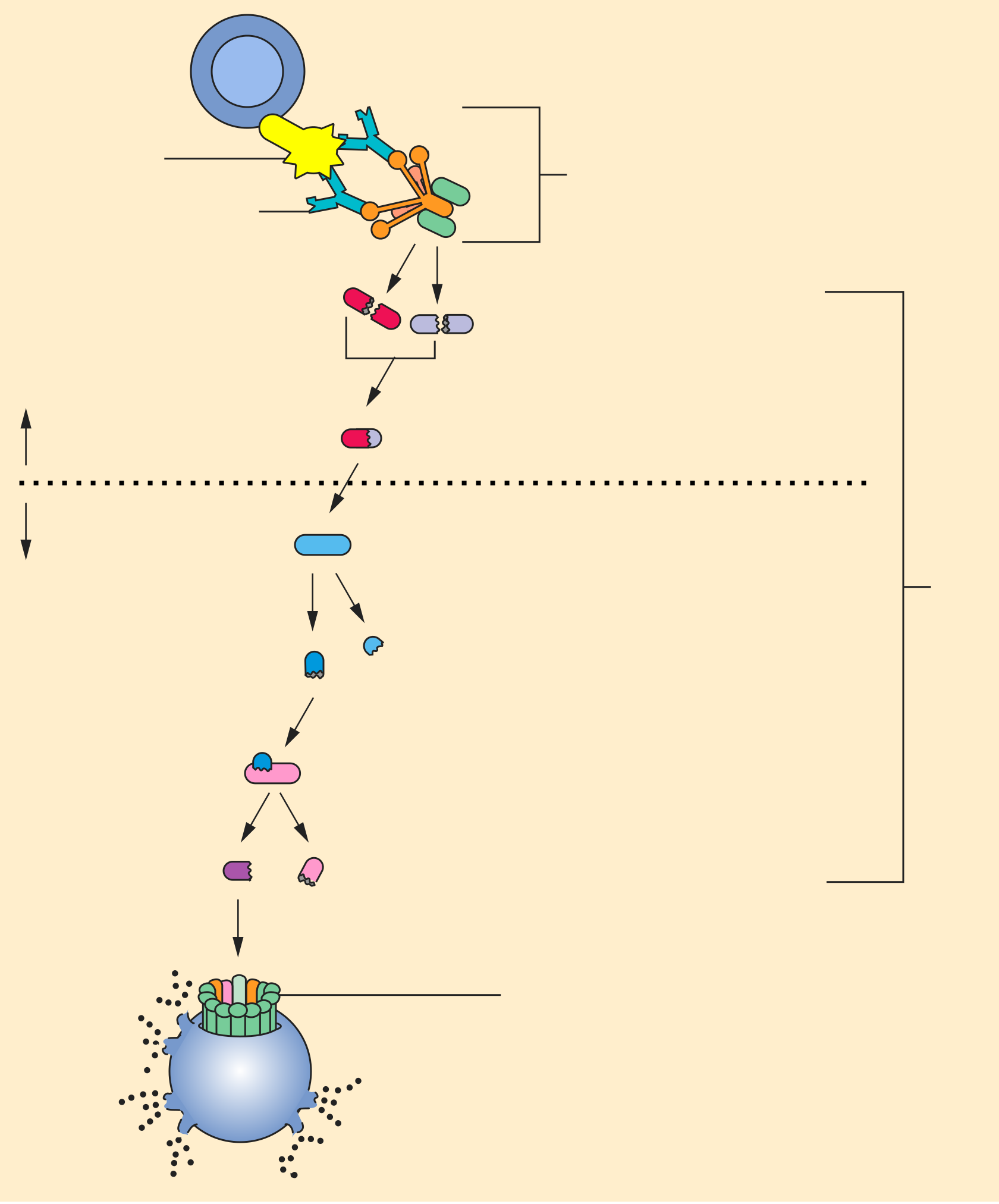
- All three pathways converge on C3 convertase → cleaves C3 → C3a + C3b; adding C3b builds C5 convertase → cleaves C5 → C5a + C5b
- Two primary opsonins = C3b and IgG (C3b binds phagocyte receptor CR1 / CD35)
- Anaphylatoxins = C3a, C4a, C5a → mast cell/basophil degranulation (histamine); C5a is the most potent
- C5a = the chemotactic fragment → recruits neutrophils
- MAC = C5b–C9 → osmotic lysis; the decisive defense against Neisseria (thin gram-negative cell wall)
- Classical = IgG or IgM immune complexes; Lectin = MBL on mannose; Alternative = microbial surfaces / LPS (spontaneous C3 tickover, stabilized by properdin)
- Convertase shorthand: classical/lectin C4b2a, alternative C3bBb
The Three Activation Pathways
| Pathway | Trigger / Activator | Initiator molecule | Board pearl |
|---|---|---|---|
| Classical | IgG or IgM bound to antigen (immune complexes) | C1 complex (C1q–C1r–C1s) | "GM makes classic cars" |
| Lectin | Mannose / carbohydrates on microbial surface | Mannose-binding lectin (MBL) + MASP | Innate; needs no antibody |
| Alternative | Microbial surfaces, LPS/endotoxin; spontaneous C3 "tickover" | C3b + Factor B, Factor D, properdin | Properdin deficiency → Neisseria |
Activation → Amplification → Lysis
The cascade is one enzymatic relay:
- Initiation differs by pathway, but all three assemble a C3 convertase (classical/lectin C4b2a; alternative C3bBb, stabilized by properdin).
- C3 convertase cleaves C3 → C3a (anaphylatoxin) + C3b (opsonin that also amplifies the loop). Deposition of another C3b converts it into C5 convertase (C4b2a3b or C3bBb3b).
- C5 convertase cleaves C5 → C5a (chemotaxis + anaphylatoxin) + C5b. C5b nucleates C6–C7–C8, then polymerized C9 forms the pore = membrane attack complex, causing osmotic lysis.
Regulation is the safety catch. Fluid-phase and GPI-anchored membrane inhibitors keep complement off host cells — DAF (CD55) and CD59 on RBCs, and C1 esterase inhibitor in plasma. Lose the regulator and self-tissue takes the hit (PNH, hereditary angioedema).

Molecule / Regulator → Function → Disease
| Molecule / regulator | Function | Deficiency or defect → disease |
|---|---|---|
| C3b | Opsonization (binds phagocyte CR1/CD35); immune-complex clearance | C3 deficiency → severe, recurrent pyogenic infections (encapsulated bacteria) + type III hypersensitivity (immune-complex disease) |
| C3a, C4a, C5a | Anaphylatoxins → mast cell/basophil histamine release | — (C5a most potent) |
| C5a | Neutrophil chemotaxis (+ anaphylatoxin) | — |
| C5b–C9 (MAC) | Membrane attack complex → lysis | Terminal (C5–C9) deficiency → recurrent Neisseria (meningitidis & gonorrhoeae) |
| C1 esterase inhibitor | Inhibits C1 and kallikrein (limits bradykinin) | Deficiency → hereditary angioedema (↑bradykinin); ↓C4; avoid ACE inhibitors |
| DAF (CD55) & CD59 | GPI-anchored: DAF decays C3 convertase; CD59 blocks MAC (protect self RBCs) | PNH (PIGA mutation → no GPI anchor) → intravascular hemolysis + thrombosis |
| Early classical: C1q, C4, C2 | Clear immune complexes & apoptotic cells | Deficiency → SLE; C2 = most common complement deficiency |
- "GM makes classic cars" — IgG and IgM activate the Classical pathway.
- Anaphylatoxins are the small "a" fragments — C3a, C4a, C5a trigger histamine; C5a is the most potent and the chemotactic one.
- "No MAC → Neisseria attack" — terminal complement (C5–C9) or eculizumab-blocked patients get recurrent Neisseria.
- "C1 INhibitor INhibits" — lose the inhibitor, release the brake on bradykinin → hereditary angioedema.
- "C3b binds bacteria" — alliteration for C3b = opsonin → phagocytosis.
- Recurrent Neisseria — 19-year-old dorm resident with a second episode of meningococcal meningitis (or recurrent disseminated gonococcal infection); CH50 is low. → Terminal complement (C5–C9) deficiency (consider properdin deficiency if the alternative-pathway screen / AH50 is abnormal but CH50 is normal).
- Hereditary angioedema — recurrent non-pitting, non-pruritic, urticaria-free swelling of lips/face/larynx and colicky bowel edema; family history; an attack follows starting an ACE inhibitor; C4 is low. Antihistamines and epinephrine don't work (it's bradykinin-mediated). → C1 esterase inhibitor deficiency.
- PNH — young adult with episodic dark (cola-colored) morning urine, fatigue, and an unexplained venous thrombosis (e.g., Budd–Chiari); flow cytometry shows loss of CD55/CD59. → Paroxysmal nocturnal hemoglobinuria; treat with eculizumab (anti-C5) and vaccinate against meningococcus first.
- SLE + infections — young woman with malar rash and arthritis plus recurrent sinopulmonary infections; homozygous early-classical (C1q/C4/C2) deficiency. C1q deficiency carries the highest single-gene risk for lupus (rare but highly penetrant).
- CH50 tests the classical pathway (needs C1–C9); a low CH50 points to a classical or terminal component deficiency.
- Low C3 AND low C4 → active SLE, serum sickness. Low C3, normal C4 → post-streptococcal GN and membranoproliferative GN (C3 nephritic factor stabilizes C3 convertase, consuming C3).
- Eculizumab = anti-C5 monoclonal antibody (blocks C5a + MAC); used in PNH and atypical HUS → creates functional terminal deficiency → ↑ Neisseria risk → give meningococcal vaccine first.
- Hereditary angioedema management: C1-INH concentrate, icatibant (bradykinin B2 antagonist), or ecallantide (kallikrein inhibitor).
- Quick recall: opsonins = C3b + IgG; chemotaxis = C5a; anaphylatoxins = C3a/C4a/C5a; MAC = C5b–C9.
Practice Immunology now
Board-style questions, spaced-repetition flashcards, and a Socratic AI tutor — free to start.